


Abstract
Epithelioid hemangioendotheliomas are rare vascular tumors, often arising from medium to large veins in the extremities. Symptoms of these tumors vary depending upon location. Rarely, tumors may arise in chest and involve large vessels in the mediastinum. We present a case of a 17-year-old male presenting with compressive symptoms of the left upper extremity who was found to have a large epithelioid hemangioendothelioma encasing the left brachiocephalic vein.
INTRODUCTION
Epithelioid hemangioendothelioma is a rare vascular neoplasm, considered to have clinical behavior intermediate between a benign hemangioma and malignant angiosarcoma [1]. The tumor arises from endothelial cells. First described in 1982 by Weiss and Enzinger, these tumors often arise from a medium-to-large-caliber vein, most commonly in the extremities. Mediastinal locations are reported in 8% of cases [2–4]. Symptoms of these tumors vary depending on location, and can present with extremity swelling, pain, cough or dyspnea. Epithelioid hemangioendotheliomas are most commonly diagnosed in adults. Tumor size and mitotic activity have been correlated with survival. Complete surgical excision of these tumors is advocated given their potential for malignant degeneration and metastasis.
CASE REPORT
A 17-year-old male presented with complaints of left-arm swelling and occasional pain. Imaging obtained of the chest and neck revealed a large anterior mediastinal mass involving the major venous structures of the left chest (Fig. 1). Computed tomography (CT)-guided needle biopsy was performed, with inconclusive results, and this was followed by a left video-assisted thoracoscopic surgery (VATS) procedure yielding enough tissue for formal diagnosis of an epithelioid hemangioendothelioma. After extensive multidisciplinary consultation and preoperative planning, the patient was taken to the operating room for a median sternotomy, thymectomy and en-bloc excision of the tumor and vessels (Fig. 2). Vascular reconstruction of the left brachiocephalic and left subclavian vein was performed with an 8 mm ringed polytetrafluoroethylene graft, with an additional end-to-side anastomosis of the left internal jugular vein to the graft. The patient tolerated the initial procedure well, was extubated on postoperative Day 1 and advanced to an oral diet. After several days, large-volume milky drainage from the chest tube was evaluated and presence of a chyle leak confirmed. The patient returned to the operating room on postoperative Day 6 for a right VATS with ligation of the thoracic duct. Final tumor measurements were 7.5 × 5.5 × 3.5 cm, and final pathology confirmed an epithelioid hemangioendothelioma with no mitoses, <5% necrosis and a final pathologic staging of pT2b. The patient has since recovered uneventfully and was discharged home in stable condition.
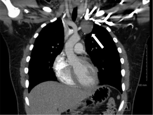
Computed tomography demonstrating a mass in the left chest involving the brachiocephalic vein.
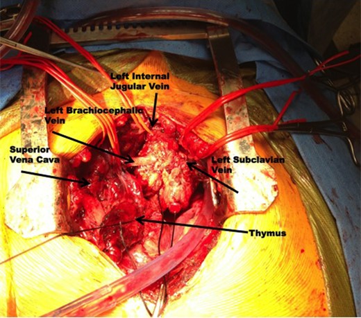
Intraoperative photography demonstrating the epithelioid hemangioendothelioma encasing the left brachiocephalic vein, left internal jugular vein, and left subclavian vein.
DISCUSSION
Epithelioid hemangioendotheliomas are unique vascular neoplasms, occurring relatively infrequently and sporadically. Malignant degeneration and metastases are reported to occur in 25% of patients, with death occurring in up to 15% of patients [5]. Despite the indolent clinical course, the potential for tumor advancement mandates complete surgical excision. The vascular involvement necessitates meticulous preoperative planning and multidisciplinary approach for aggressive resection and concomitant reconstruction of involved vessels. In this case, the surgical team included cardiothoracic surgeons, vascular surgeons and a pediatric surgeon.


