


Abstract
Vaginal vault dehiscence (VVD) may occur rarely after hysterectomy. Although mostly, a vaginal cuff dehiscence is seen after robotic or laparoscopic hysterectomy, it may also be observed as a complication of abdominal or vaginal hysterectomy. Vaginal repair is one of the techniques used for VVD. Here, we will describe a case of vaginally repaired VVD, associated with intra-abdominal hematoma after postpartum hysterectomy.
INTRODUCTION
Vaginal vault dehiscence (VVD) is a rare and serious complication of hysterectomy. It may cause mortality and morbidity [1]. Incidence is low and varies between 0.03% and 0.28% [2]. Mode of hysterectomy affects the incidence. Although the incidence of VVD is below 1% in overall hysterectomies, recent data have indicated that the incidence may be as high as 4% after conventional laparoscopic and robotic laparoscopic surgery [2, 3]. Patient characteristics like smoking, diabetes, chronic lung disease increasing intra-abdominal pressure by coughing and constipation, malignity, vaginal atrophy, advanced age, chronic steroid therapy, postoperative radiation therapy and additional risk factors like early sexual intercourse, postoperative cuff infection and hematoma are associated with VVD [4, 5].
Here, a case of VVD associated with intra-abdominal hematoma after postpartum hysterectomy repaired by vaginal suturing technique will be discussed.
CASE PRESENTATION
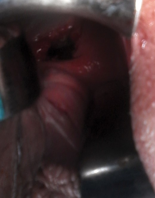
A 32 year old woman, G8 P2 A5, 39 weeks of gestation was referred to obstetric clinic of Adnan Menderes University, School of Medicine with complaint of vaginal bleeding. Total placenta previa was diagnosed by ultrasonography and magnetic resonance imaging (MRI). She underwent an emergency caesarian section. A 2450-gr baby was born with 1 and 5 min. APGAR score of 8/9. Placenta could not be removed completely as it invaded in right-posterior wall of lower segment of uterus. Postpartum hysterectomy was performed to stop bleeding. The vaginal vault was closed with polyglactin 910 (Vicryl-1/Ethicon) continuous interlocking suture. The estimated blood loss was over 2 L. She was administered 4 U of packed cells, fresh frozen plasma, and she was observed on the intensive care unit in the postoperative period for two days. Postoperative recovery was uneventful, and she was discharged 5 days after the operation. On day 21, she applied to emergency clinic by heavy vaginal bleeding and watery discharge. An intra-abdominal hematoma of approximately 16 × 10 × 9 cm in diameter was observed in ultrasonography and MRI. In pelvic examination, vaginal cuff dehiscence was observed (Fig. 1). The patient was hemodynamically stable. Intravenous broad spectrum antibiotic had been started. On day 2 of the admission, exploratory laparotomy was performed. Dense adhesions between bowel and bladder were observed. Intra-abdominal hematoma was located between these dense adhesions of bowel, on vaginal vault which could be hardly evaluated. Hematoma was drained abdominally. It was impossible to explore vaginal vault by abdominal surgery because of the dense adhesions. Operation continued in lithotomy and Trendelenburg position. Examination of the vaginal vault in lithotomy position showed the entire dehiscence. Necrotic tissue was debrided, and irrigation, aspiration was performed. Vaginal cuff was closed with synthetic absorbable suture using interrupted sutures. Her postoperative recovery was uneventful and she was discharged on day 29.
Vaginal Vault Dehiscence associated with intra-abdominal hematoma after postpartum hysterectomy.
DISCUSSION
Although rare, VVD can represent an extremely rare serious postoperative complication of hysterectomy and may cause morbidity for patients. Although it is more commonly seen in laparoscopic and robotic hysterectomy [6], it can be also seen in abdominal and vaginal route as in the current case. It is mostly seen in three months after hysterectomy, occurring earlier in endoscopic route [7]. Interestingly, it occurred on day 21 of operation in our case and risk factors are blamed for early development of VVD.
A number of different possible risk factors have been proposed in the published data. Nick et al. [8] conducted a study to determine the vaginal cuff separation and associated risk factors in patients undergoing laparoscopic and robotic hysterectomy. They reported that, sexual intercourse in 8–48% and defecation or Valsalva testing (cough or sneeze) in 16–30% of cases as the precipitating event. However, spontaneous vaginal cuff dehiscence was reported to represent up to 70% of the cases. Increased age, increased number of vaginal surgeries, vaginal atrophy, factors that are associated with poor wound healing (including malignancy, chronic steroid use, malnutrition, tissue radiation), and postoperative vaginal cuff infection or hematoma may be other risk factors [9]. In the current case, postoperative hematoma was main risk factor. In addition, our patient was a heavy smoker and had chronic cough due to it.
Many conditions in the operation technique play role in development of vaginal cuff dehiscence. First of all, mode of hysterectomy affects the incidence of dehiscence. Moreover, technique of colpotomy at hysterectomy has been associated with cuff breakdown. In laparoscopic surgery, vaginal tissue damage attributable to monopolar energy used in colpotomy and bipolar cauterization for hemostasis is blamed. It was speculated that the cauterization with consequent cell necrosis and tissue damage associated with the use of monopolar knife could have a negative effect on the healing of the vaginal cuff. Moreover, bipolar energy used for hemostasis cause greater tissue damage. So, monopolar energy modality with low voltage causing minimal tissue damage is advised for reducing VVD [10]. In this patient, cold knife colpotomy was performed as used in most abdominal and vaginal hysterectomies.
In the present case, it is more likely that a VVD occur secondary to an underlying factor such as a hematoma or a primary healing defect as a result of excessive coagulation. Hypothetically, vaginal wall epithelium remains approximated only by the suture. Therefore, as soon as the suture loses most of its tensile strength, a (partial) separation of the vaginal cuff occurs. This hypothesis is supported association of intra-abdominal hematoma and VVD, as we found in the present case. Hematoma increases the intra-abdominal pressure by its mechanical effect, and also causes pressure necrosis on tissue. Both these pathways decrease strength of sutures and VVD occurs. So, emergent surgical intervention is warranted. In VVD, a surgical intervention is mandatory and prompt repair is advisable. There is no consensus about the ideal method of surgical repair.
Best comment for VVD is to prevent its occurrence. Besides all known risk factors about it, we would like to emphasize importance of hemostasis in hysterectomy. All precautions should be taken to prevent complications especially hematoma. In conclusion, we advice to drain intra-abdominal hematoma as early as possible as it seems to be a major risk factor for vaginal cuff dehiscence.
CONFLICT OF INTEREST STATEMENT
The authors have no conflicts of interest to declare.
