


Abstract
Augmentation mammoplasty is the most common aesthetic surgical procedure performed in the USA. Prosthetic failure is a major reason for surgical reintervention. A number of causes for this have been documented, but costal bone abnormalities leading to perforation of the prosthesis are very unusual. We present the case of a woman who experienced spontaneous deflation of both saline implants in close succession, and who was found to have sharp bony spicules on both sides of her chest. Pathology examination reported reactive changes, suggestive of heterotopic bone. Examination of the implants showed no defects besides small punctures on the back wall, which coincided with the position of the spicules of bone. There are a number of possible causes for these bony growths which we examine in turn. The chest wall should be examined in all cases where unexplained implant deflation has occurred.
INTRODUCTION
Augmentation mammoplasty is the most common aesthetic surgical procedure performed in the USA [1]. The incidence of secondary surgery is as high as 36% [1]; a significant proportion was due to deflation. For saline implants, the incidence of deflation was 8.3% over a mean patient follow-up period of 13 years; the vast majority of which were spontaneous [2]. Reasons for this include fold defects [3] and under-filling, though this has been questioned [2]. Implant deflation due to anatomical abnormalities has been documented very rarely. We present the case of a 42-year-old woman who experienced deflation of both of her saline implants ∼15 months apart. Examination of her chest wall after implant removal showed sharp bony projections on both sides.
CASE REPORT
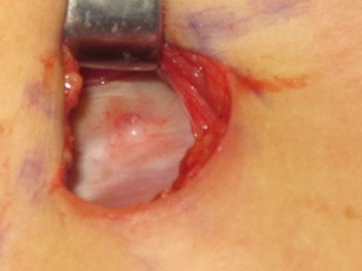
The spur of bone on the right chest wall of the 42-year-old patient immediately after removal of the deflated implant.
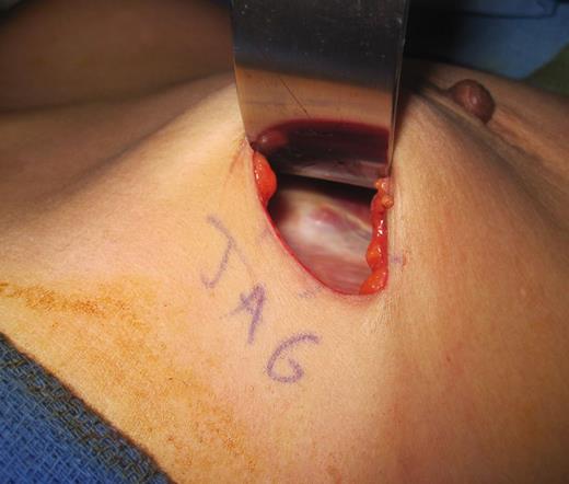
The bone spicule similar to that found on the right but this time on the left chest wall immediately after implant removal.
Gross examination of both deflated implants revealed no defects except for single incisions on the back wall, which coincided with the position of the bony prominences described.
DISCUSSION
The precise origin of these spicules is unclear and there is probably a combination of factors responsible. This combination of predisposing factors may explain why the complication is so rare.
The previous report of costal exostosis leading to prosthetic rupture proposes injury or trauma to the ribs as a possible explanation [4]. Chronic stress from flexion and rotation of the thoracic cage has also been associated with hyperostosis [5]. Osteophytes have been implicated in thoracic pathology including hemothorax [4] and recurrent pleural effusions [6]. Osteophyte formation has a significant individual variation in growth in response to trauma and stress [7]. It may be that the patient had a genetic predisposition to form these bony processes in response to injury or chronic stress on the rib cage. Placing the original prostheses in the subpectoral plane may have injured the chest wall. Additionally, the presence of the implant itself may have placed the thoracic cage under non-physiological stresses, which led over years to the formation of bony spurs.
Heterotopic ossification in the chest has been reported [8]. It involves bone growth in soft tissues and can have a range of etiologies, including post traumatic [9]. This is self-limiting and involves a local proliferation of fibroblasts and bone formation in skeletal muscle. Mature bone and cartilage are present, without cytological atypia, as was found in our patient [9]. Heterotopic bone formation is thought to involve a number of factors including signaling abnormalities, such as overexpression of bone morphogenic proteins and under-expression of their antagonists [9]. Abnormalities such as these are thought to be the most crucial to heterotopic bone formation [9]. It is not known if the patient had heterotopic ossifications elsewhere in her body.
The prevention of heterotopic ossification and its recurrence is challenging. A number of effective methods exist including long-term non-steroidal anti-inflammatory drug therapies and radiotherapy [10]. However, these carry significant side effects. Bone morphogenic protein receptor antagonists are being tested [10].
Capsular contraction may also play a role in the presence of bone spicules [4], with the formation of a tight collagen capsule increasing compression.
Interestingly, the patient in our case was of an Asian ethnicity and the only similar report of this event [4] occurred in Taiwan. While no firm conclusions can be drawn from only two individuals, it may be possible that Asian recipients of augmentation mammoplasty are more prone to this complication.
In conclusion, multiple factors including genetic predisposition, possible surgical trauma and capsular contraction may be collectively responsible for this complication and this possibly explains why it occurs so rarely. Both prostheses failed resulting in two further operations. Placing new implants without removing the sharp spurs of bone would have left the patient with a significant chance of experiencing further deflations. Surgeons should be aware of this possible complication, and the chest wall should always be examined after unexplained prosthetic deflation.
REFERENCES
Author notes
This paper has not been presented at any meeting.
