-
PDF
- Split View
-
Views
-
Cite
Cite
Adarsh Vijay, Husham Abdelrahman, Ayman El-Menyar, Hassan Al-Thani, Early laparoscopic approach to pancreatic injury following blunt abdominal trauma, Journal of Surgical Case Reports, Volume 2014, Issue 12, 1 December 2014, rju129, https://doi.org/10.1093/jscr/rju129
Close - Share Icon Share
Abstract
The incidence of pancreatic injury following blunt abdominal trauma is rare. A timely accurate diagnosis of such injury is difficult and also the management remains controversial. Here, we reported the successful use of laparoscopy to diagnose, characterize and treat blunt pancreatic trauma in a 28-year-old male patient involved in a motor vehicle crash. An abdominal computed tomography scan showed peripancreatic fat stranding suggestive of pancreatic injury. With persistent clinical signs of peritonitis and laboratory investigations suggestive of pancreatitis, the patient underwent laparoscopic drainage of the lesser sac. The patient had an uneventful postoperative course. The management of patients with blunt pancreatic injuries should be tailored to individual situations. Our experience suggests that a timely laparoscopic management of traumatic pancreatic injury is safe approach in selected cases.
INTRODUCTION
Owing to its protected retroperitoneal location, pancreatic injury following blunt trauma is rare, with an incidence rate of <2% [1]. Blunt abdominal trauma accounts for 20% of all cases of traumatic pancreatitis [2]. Early and accurate identification of such injuries are imperative to avoid high morbidity and mortality associated with ductal injuries. A conservative management approach has been advocated when a ductal injury is not evident. However, proponents of early surgical intervention attributed to the delay in intervention to be related with increased rates of morbidity and mortality [1–3]. So, an early laparoscopic approach might play a role for the diagnosis and management of blunt pancreatic injuries. Here, we report a case of traumatic pancreatitis following blunt abdominal trauma managed successfully using laparoscopic techniques.
CASE REPORT

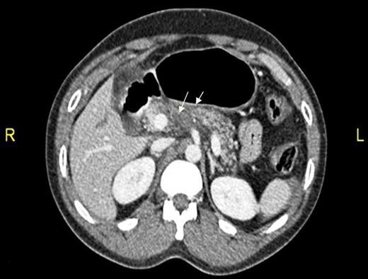
CT abdomen showing two hepatic lacerations in addition to minimal subhepatic and left subdiaphragmatic free fluid.

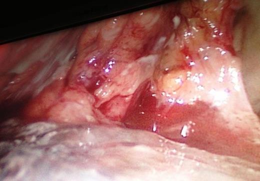
The intraoperative extension of the pancreatic necrosis that involves the entire gland.

A 6-month out-patient abdominal CT follow-up showed no fluid collection and the patient was asymptomatic.
DISCUSSION
Definite management of pancreatic injuries is not well defined yet. Moreover, the clinical and laboratory findings of blunt pancreatic injury are nonspecific, and therefore accurate diagnosis in its acute phase is usually delayed [1, 3]. Computed tomography scanning may fail to delineate the pathology, especially in the initial stage. Also, the ability for CT to demonstrate the integrity of pancreatic duct is limited (43%); however, multiple detector computed tomography may have better result [1, 4, 5].
MRCP allows direct imaging of the pancreatic duct and its disruption [1]. ERCP seems the most reliable diagnostic tool to accurately define the continuity of the main pancreatic duct following pancreatic trauma with a 100% sensitivity and specificity [6]. However, the need for a skilled endoscopist and the invasive nature of procedure may limit its use in unstable patients. Suspicion of concomitant traumatic intra-abdominal injuries prompts the surgeon to choose exploration over ERCP. Pancreatic lacerations not involving the duct (The American Association of Surgeons in Trauma Grade I and II) are considered minor injuries and are predominantly managed conservatively. Grade III–V injuries usually necessitate surgical exploration depending on the degree of injury and its proximity to the mesenteric vessels; where distal pancreatectomy is the most common approach [7]. Kantharia et al. [8] studied 17 cases with pancreatic trauma and found that contrast-enhanced CT as the useful tool for diagnosis and grading of the pancreatic injuries. Moreover, the majority of these cases responded well to the conservative treatment; however, higher grade pancreatic injury required surgical intervention but with higher mortality.
In patients with blunt pancreatic trauma, delayed morbidities include pancreatitis, fistula, abscess, pseudocysts and major duct strictures [9]. Therefore, we assume a valid role for early laparoscopic approach for suspected injuries. This is particularly compelling when CT scan is inconclusive and patients show signs suggestive of peritonism. Intraoperative diagnosis of ductal injuries is seldom done owing to its complexity and retrograde pancreatography through a small duct is often difficult in trauma patients.
Our patient's clinical deterioration precluded the use of preoperative ERCP/MRCP. Clinical suspicion for hollow viscus injury prompted diagnostic laparoscopy. Necrotic and hemorrhagic pancreas precluded any intraoperative attempt to accurately characterize extent of ductal injury. Looking retrospectively, we acknowledge that, if we were able to diagnose ductal transection pre/intraoperatively, our management would have drastically deferred towards distal pancreatectomy.
In addition to being an exploratory, laparoscopy can be effectively used to characterize pancreatic injuries (grading) that informs the management with immediate drainage for the proximal pancreatic part or the resection of the distal injuries, whenever indicated [10]. Therefore, a laparoscopic approach provides a genuine alternative to the open abdominal surgery.
In conclusion, the management of blunt pancreatic injuries should be tailored to individual situations. In selected patients with worsening symptoms, a prompt trial of laparoscopy is an useful option. Although we chose a conservative drainage approach due to inconclusiveness on the integrity of pancreatic duct, we were able to achieve a favorable result. Further studies are needed to confirm our findings.
AUTHORS' CONTRIBUTIONS
A.V. was involved in study design, data collection and writing manuscript. H.A.: data analysis and interpretation, drafting and manuscript review; A.E.M.: data analysis and interpretation, drafting and manuscript review; H.A.T.: study design, data interpretation and review manuscript.
CONFLICT OF INTEREST STATEMENT
None declared.
ACKNOWLEDGEMENTS
The authors thank all the trauma surgery staff for their cooperation. All authors read and approved the case report. This case report has been approved by the Medical Research Center (IRB no. 13311/13), Hamad Medical Corporation, Doha, Qatar.


