


Abstract
Transverse testicular ectopia (TTE) is a rare anomaly that is commonly associated with inguinal hernia. Most of the reported cases are in children with very few reported cases in adults. We report a case of 42 years, fertile male, who presented with left reducible inguinal hernia. During surgery, he was found to have a left indirect inguinal hernia with TTE with both testes on the left side. Hernioplasty and bilateral orchidopexy were performed. He had an uneventful recovery. Most of these cases are diagnosed intraoperatively, but imaging (ultrasonography and magnetic resonance imaging) has emerged as a promising tool for preoperative diagnosis although ultrasound missed it in this case.
INTRODUCTION
Transverse testicular ectopia (TTE) is an uncommon anatomical abnormality in which both the gonads migrate towards the same hemiscrotum. The ectopic testis may lie in the opposite hemiscrotum, in the inguinal canal or at the deep inguinal ring. An inguinal hernia is commonly present on the side to which the ectopic testis has migrated [1]. The diagnosis is commonly established during surgical exploration.
Here, we present a case of 42-year-old male diagnosed preoperatively as left inguinal hernia with normal ultrasonography findings, who found to have TTE on exploration.
CASE REPORT
A 42-year-old married male, having three children, presented to us with history of swelling over left inguinal region for 2 years. The swelling was painless, gradually increasing in size and more prominent on coughing and straining. He did not have chronic cough, constipation or urinary complaints. On examination, there was a 3 × 3 cm reducible left indirect inguinal hernia. An ultrasonography revealed a normal scan of bilateral testes, scrotum and inguinal region. He was planned for hernioplasty and on exploration, there was a left indirect inguinal hernia with thinned out hernial sac with vas and cord structures of the left side. Surprisingly, both testes could be delivered into the wound with their individual vas deferens and cord structures (Fig. 1). Both the cord structures were passing through the internal ring of the left side. Both of them were mobilized, followed by right orchidopexy (of medially located testis) by passing it through a trans-septal incision. Left orchidopexy was also performed. Herniotomy and mesh repair were performed on the left side. He had an uneventful recovery and on follow-up, he had no complications; both the testes were normal to feel with no vascular compromise on Doppler study. He is on a regular follow-up with no complications till date.
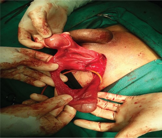
Exploration of left inguinal region revealed both testes with cord structures on the left side.
DISCUSSION
TTE, also called testicular pseudoduplication, unilateral double testis and transverse aberrant testicular maldescent, is an uncommon anatomical abnormality in which both the gonads migrate towards the same hemiscrotum. It was first reported by Von Lenhossek in 1886 [2], as an autopsy finding. More than 100 cases have been reported in the literature. The embryological etiology of TTE is controversial. Adhesion or fusion of developing Wolffian ducts, defective or aberrant gubernaculum, testicular adhesion, defective formation of the internal inguinal ring, traction on a testis by persistent Müllerian structures and possibility of the development of both testes from the same germinal ridge, are some of the postulated theories for the ectopic testis. Association of TTE with persistent Müllerian ducts was first described in 1895 by Jordan [3]. Mechanical effect of persistent Müllerian duct structures may prevent the testicular descent or lead to both testicles descending towards the same hemiscrotum, producing TTE. TTE may have an increased risk of malignancy as any other forms of ectopic testis or undescended testis, so long-term follow-up is required [4].
TTE is classified into three types based on associated abnormalities [5]:
Type I: Accompanied only by hernia (40–50%).
Type II: Accompanied by persistent or rudimentary Müllerian duct structures (30%).
Type III: Associated with disorders other than Müllerian remnants, e.g. hypospadias, true or pseudohermaphroditism and other scrotal abnormalities (20%).
Patients with TTE commonly present as inguinal hernia on one side and absent testis on the other side. Most cases of TTE are diagnosed intraoperatively [6]. In our case, the patient had left inguinal hernia and absent testis on the right hemiscrotum, which we could not identify on clinical examination and ultrasonography. One explanation could be that since both the testes were on the left hemiscrotum, the pressure effect of left testis on right one could have pushed the right testis towards further right of the left hemiscrotum giving a false impression on clinical examination that location of right testis was on correct side. There are reports suggesting ultrasonography, CT scan, magnetic resonance imaging and magnetic resonance venography as tools for preoperative diagnosis of TTE [7]. Few cases of TTE in adults have been reported [8, 9].
The case presented here was managed by orchidopexy of the correctly lateralized testis to the ipsilateral hemiscrotum, and orchidopexy of the crossed testis to the contralateral hemiscrotum through a trans-septal incision, known as the Ombredanne procedure [4].
This case is reported with a view that surgeons need to be aware of this anomaly during repair of inguinal hernia, as most cases of TTE are diagnosed intraoperatively, hence adequately and safely treat the patient even with TTE when discovered unexpectedly, continue long-term follow-up to identify malignancy early, if any. Patients with TTE need to be treated by restoring the contralateral testis to its original hemiscrotum through a trans-septal incision. Written informed consent was obtained from the patient for publication of this case report and related photograph.
AUTHORS' CONTRIBUTIONS
All three authors were involved in the treatment of the patient and wrote and finalized the manuscript.
CONFLICT OF INTEREST STATEMENT
The authors declare that they have no competing interests.
