-
PDF
- Split View
-
Views
-
Cite
Cite
Jonathan Green, Luke G. Gutwein, Amyand's hernia: a rare inguinal hernia, Journal of Surgical Case Reports, Volume 2013, Issue 9, September 2013, rjt043, https://doi.org/10.1093/jscr/rjt043
Close - Share Icon Share
Abstract
Inguinal hernia repair is commonplace in general surgery practice and an estimated 700 000 are performed each year in the USA. The presence of the vermiform appendix contained in the hernia sac, or an Amyand's hernia, is exceedingly rare, occurring in 1% of inguinal hernia patients. We report the intra-operative findings of a standard inguinal hernia repair and discuss the management of the four types of Amyand's hernia.
INTRODUCTION
An Amyand's hernia is a rare occurrence where the vermiform appendix is found in an inguinal hernia sac. It is most commonly found intra-operatively during a right-sided inguinal hernia repair [1]. We present a case in which an Amyand's hernia was discovered in a patient with both an umbilical and a right-sided inguinal hernia.
CASE REPORT
A 61-year-old African-American male was seen in the surgery clinic with a 6-year history of an umbilical and enlarging right inguinal hernia. The umbilical hernia was symptomatic causing intermittent discomfort. The right inguinal hernia was also symptomatic and had progressively enlarged over time. He denied any changes in bowel habits or history of intestinal obstruction.
On physical examination, his abdomen was soft, non-tender and non-distended. A small supra-umbilical tender bulge was present and incarcerated. An inguinal examination was significant for a right-sided, tender, reducible mass without scrotal involvement.
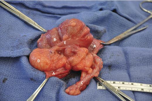
On the day of surgery, the patient was prepped, and draped in an aseptic technique. Initially, the umbilical hernia was repaired primarily without complication. The right inguinal hernia repair was approached with a 5 cm right-sided oblique incision parallel to the inguinal ligament. Subcutaneous tissue through Scarpa's fascia was divided until aponeurotic fibers of the external oblique were visualized. After dividing the external oblique to the superficial inguinal ring, the contents of the inguinal canal were then circumscribed using blunt dissection. The hernia sac lateral to the inferior epigastric pedicle was dissected away from the spermatic cord to the deep inguinal ring. The sac was opened illustrating the cecum and vermiform appendix (Fig. 1). There were no inflammatory changes of the appendix or cecum. The sac contents were reduced into the peritoneal cavity. The hernia sac was excised and the peritoneum was suture ligated. We performed a tension-free polypropylene mesh repair. The patient was discharged the same day. He returned to clinic 1 month later with no complications and no recurrence of his hernias.

Hernia sac (A) opened containing cecum (B) and vermiform appendix (C); epiploic appendage (D), tenia coli (E).
DISCUSSION
Claudius Amyand was a French born English Surgeon who in 1735 successfully performed and recorded the repair of an inguinal hernia in an 11-year-old patient. The patient was found to have the vermiform appendix in his hernia sac. Since then, the presence of the vermiform appendix in a hernia sac has been deemed an ‘Amyand's hernia’ [2].
The incidence of an Amyand's Hernia is ∼1% of inguinal hernias occurring most often in male patients. They are most commonly located on the right side due to the location of the appendix. The appendix has also been found in obturator, umbilical and incisional hernias [1]. Of inguinal hernias, only 0.1% has an inflamed appendix [3–6]. This is a result of either primary inflammation of the appendix causing edema of the internal inguinal ring or incarceration of a normal appendix by abdominal wall musculature [1].
As in our patient, most Amyand's hernia's are discovered intra-operatively. However, pre-operative diagnosis can be made using CT with oral contrast, in patients with suspicion of appendicitis. When scrotal involvement is suspected ultrasound is a low cost alternative without radiation [1].
Losanoff and Basson created a classification scale to identify and treat Amyand's hernias (Table 1) [4, 5]. A Type 1 hernia has a normal appendix in an inguinal hernia, which is managed with a reduction and mesh repair. Types 2–4 have acute appendicitis within an inguinal hernia sac. Type 2 has an inflamed nonperforated appendix. Type 3 has a perforated appendix and type 4 is complicated with intra-abdominal pathology. Type 2–4 hernias are managed with appendectomy and primary repair (without mesh). In addition, to the primary repair and appendectomy, type 3 includes a laparotomy for abdominal irrigation, possible orchiectomy or colectomy and type 4 includes investigation of pathology [4, 5].
| Classification . | Description . | Surgical management . |
|---|---|---|
| Type 1 | Normal appendix in an inguinal hernia | Hernia reduction, mesh repair |
| Type 2 | Acute appendicitis in an inguinal hernia, without abdominal sepsis | Appendectomy, primary repair of hernia without mesh |
| Type 3 | Acute appendicitis in an inguinal hernia, with abdominal wall or peritoneal sepsis | Laparotomy, appendectomy, primary repair without mesh |
| Type 4 | Acute appendicitis in an inguinal hernia, with abdominal pathology | Manage as Type 1–3, investigate pathology as needed |
Our patient had a Type 1 Amyand's hernia and underwent a mesh repair without an appendectomy. In the pediatric population, however, a prophylactic appendectomy would have been performed (without mesh repair), because children and adolescents have a higher risk of acquiring acute appendicitis [4, 5].
In summary, Amyand's hernia is a rare occurrence, but offer variety in their presentations and managements. Our case of a Type 1 Amyand's hernia was repaired with mesh and did not require an appendectomy.


