


Abstract
Vesicoureteral reflux (VUR) affects ∼1% of children. We present an unusual case of urinary retention secondary to an obstructing urethral stone, underlying reflux, and its management. A 7-year-old boy presenting with acute urinary retention had a palpable penile shaft swelling and patent urethral meatus on examination. Cysto-urethroscopy with a 6.6Fr ureteroscope, due to unavailability of paediatric instruments, revealed an obstructing calculus impacted in the navicular fossa. This was laser fragmented and extracted. Cystoscopy revealed multiple bladder calculi with a patulous right ureteric orifice. Post-operative investigations revealed a small, scarred right kidney (ultrasound), bilateral ureteric reflux (micturating-cystourethrogram), 4 cm by 0.8 cm right ureteric calculus (CT-KUB) and 4% right split renal function (DMSA). Right laparoscopic nephroureterectomy was subsequently performed. Our case highlights the variety with which VUR can present and the effectiveness of a ureteroscope in an emergency setting as an alternative to a paediatric cystoscope to visualize the urethra and the bladder.
INTRODUCTION
Vesicoureteral reflux (VUR) affects ∼1% of children [1]. It can cause urinary tract infection (UTI), renal scarring, hypertension and renal failure. Successful management of VUR involves preventing these sequelae. Urolithiasis is a far less commonly cited consequence of VUR. We present an unusual case of a child with urinary retention secondary to an obstructing urethral stone, underlying VUR, and its acute management.
CASE REPORT
A 7-year-old boy who had no relevant past medical history (UTIs or other) presented with acute urinary retention. He was systemically well but had mild suprapubic discomfort and a palpable bladder. Examination of the penile shaft revealed a palpable hard swelling. The urethral meatus was patent. Following failure of conservative management of urinary retention (placing the boy in a warm-bath, adequate analgesia), a cysto-urethroscopy was performed under general anaesthetic. Due to lack of paediatric instruments on site, a 6.6-Fr adult ureteroscope was used. This revealed an obstructing urethral calculus impacted in the navicular fossa. The stone was fragmented using a HoYag laser and extracted. Cystoscopy with the same instrument then revealed multiple bladder calculi with a patulous right ureteric orifice. These stones were washed out and the patient was catheterized (6-Fr urethral catheter). Serum calcium, urate and renal function were normal. Post-operative ultrasound revealed a small, scarred right kidney and large (hypertrophied) left kidney. Micturating cystourethrogram demonstrated grade 1 and 2 VUR in the right and left ureters, respectively, and an opacification in the upper right ureter suspicious of a calculus. CT KUB confirmed a 4 cm (cranio-caudal) by 0.8cm (transverse) right ureteric calculus (Fig. 1). DMSA scan revealed 4% (right) split renal function. The patient subsequently underwent a right laparoscopic nephroureterectomy (Fig. 2).
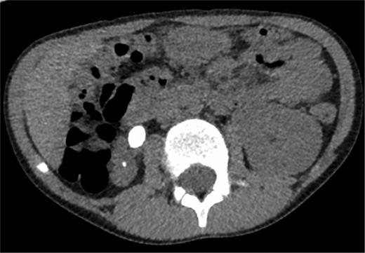
Non-contrast CT demonstrating the large right ureteric calculus.
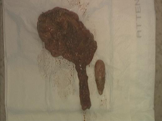
Right kidney and ureter with calculus.
DISCUSSION
VUR is diagnosed most commonly in children presenting with UTI. However, its predisposition to stone formation is the process that leads to our patient's presentation. Children with VUR are more likely to have hyper calciuria and hyper uricosuria, as well as UTIs and urinary stasis, which promote urinary crystal and subsequent stone formation. Studies show that the estimated prevalence of VUR in patients with urolithiasis is between 8 and 18% [2]. Although girls are more commonly diagnosed with VUR (due to the higher incidence of UTIs), boys often have a higher grade of reflux. In this case, the right ureteric reflux was graded 1 radiologically, but was likely to be an underestimate due to the presence of an impacted ureteric calculus preventing higher grade reflux. Spontaneous resolution of VUR depends on the grade. Studies suggest nearly 80% of Grade 1–2, and 30–50% of Grade 3–5 reflux will resolve based on 5-year follow-up data [3, 4]. Bilateral reflux is reported to take longer to resolve than unilateral reflux [5].
Management of reflux is broadly divided into medical versus surgical. As they are highly likely to resolve spontaneously, grades 1–3 are generally managed non-operatively with preventing UTIs until resolution of reflux. Controversy, however, exists over the use of routine prophylactic antibiotics in low-grade reflux. Whereas earlier studies found continuous antibiotic prophylaxis useful at reducing the risk of febrile UTIs, recent studies have not mirrored this conclusion and raise the issue of antibiotic resistance [6]. In our case, the young boy has been commenced on continuous antibiotic prophylaxis after careful discussion with parents regarding the risks and benefits of this approach in light of the limited literature available. He will be followed up with annual ultrasound to monitor his remaining left kidney.
Recurrent UTIs resistant to antibiotic therapy and breakthrough infections in those on continuous prophylactic antibiotics are indications for intervention. Options available are endoscopic injection (first reported in 1984) of a peri-ureteral bulking agent and ureteral re-implantation (traditionally done open, but advances have now been made in laporoscopic technique) to achieve a submucosal length-to-width ratio of 5:1 [1].
Rigid paediatric cystoscopes range from 4.5Fr (2.4Fr working channel) to 12Fr. A newborn male urethral meatus is roughly 7Fr and increases in size with age. By age 3, over 80% of males will have a 10Fr calibre urethral meatus [7]. The use of an adult ureteroscope (6.6Fr in our case) in the absence of a paediatric cystoscope would be a reasonable alternative when urethral and bladder access is required. Our patient was certainly adequately managed in this manner. To our knowledge, there is no published literature on experience with using adult ureteroscopes for paediatric cystoscopy.
Our case highlights the variety with which VUR can present, illustrating its potential effects even in the absence of a clinical history of recurrent UTIs. Furthermore, it exhibits the importance of a thorough examination to look for rarer causes of urinary retention in the paediatric population. Lastly, the use of an adult ureteroscope in an emergency setting is an effective alternative to a paediatric cystoscope to visualize the urethra and bladder.
