-
PDF
- Split View
-
Views
-
Cite
Cite
Anne K. Marsh, Martin Hoejgaard, Incarcerated and perforated stomach found in parastomal hernia: a case of a stomach in a parastomal hernia and subsequent strangulation-induced necrosis and perforation, Journal of Surgical Case Reports, Volume 2013, Issue 4, April 2013, rjt029, https://doi.org/10.1093/jscr/rjt029
Close - Share Icon Share
Abstract
Parastomal hernias (PSHs) are a common type of incisional hernia and the most frequent complication to colostomies. Usually only mobile structures of the abdomen herniate in the hernial sac of the non-traumatic hernia. This case describes a large PSH adjacent to a lower left quadrant colostomy containing the mobile small intestine, part of the colon and a perforated stomach. The PSH presented with acute abdomen requiring explorative laparatomy and debridement. Large hernias may over time predispose to stretching of ligaments and mobilization of otherwise immobile structures with damage to these structures. The case report includes a short overview of hernia types associated with dislocation of the fixed organs of the abdominal space.
INTRODUCTION
A parastomal hernia (PSH) is a type of incisional hernia occurring at the site of the stoma or immediately adjacent to the stoma. It forms when the abdominal wall is continually stretched by the tangential forces applied to the circumference of the abdominal wall opening [1].
PSH is the most frequent complication following the construction of a colostomy. A literature review found that PSHs occur in 4.0–48.1% of patients with end colostomies [2].
Patient characteristics associated with increased risk of PSH include obesity, weight gain after ostomy construction, poor nutritional status, immunosuppressive drugs, emergency construction of the stoma, chronic coughing, infection and underlying diseases such as malignancy.
Most patients with PSH are asymptomatic and do not require surgical repair. They typically present with a bulge at the site of, or adjacent to, the intestinal stoma with or without pain. Symptoms range from mild abdominal discomfort, distension, nausea, constipation to severe abdominal pain, fever and an irreducible hernia [3].
The hernia contents are almost exclusively limited to the mobile structures of the abdomen, i.e. intestines and their supportive tissue.
This case illustrates that other contents of the abdomen may herniate as well.
CASE REPORT
An 81-year-old male patient was admitted to a surgical ward with acute onset of severe abdominal pain, vomiting and abdominal distension. Clinical examination found circulatory instability, a peritoneal abdomen and increased leucocyte count and C-reactive protein on laboratory reports, with several litres of fluid aspirated after insertion of a nasogastric tube.
His past medical record included rectal cancer 19 years ago managed with rectal resection and subsequently fitted left lower quadrant colostomy. He had developed a large PSH over a period of several years. Due to the patients’ reluctance to seek medical attention for the hernia, its diameter had increased to ∼20 cm at the time of diagnosis. Surgical correction had been deemed infeasible and a truss was fitted instead to the patient's satisfaction.
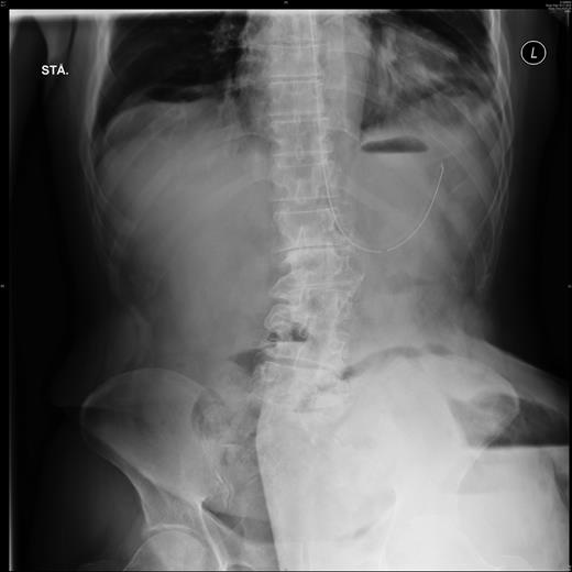
Plain film abdominal x-ray showed not only a large part of the small intestine, but also the stomach located in the PSH with signs of pneumoperitoneum (Fig. 1).

Plain film abdominal X-ray showing the trapped stomach in the large parastomal hernia on the patient's lower left side. Notice the fundus-like air on the outer left-hand side of the trapped part. Also visible is free air inside the parastomal hernia.
Acute, explorative laparotomy confirmed the finding of displacement of the stomach to the PSH with a 7 cm perforation of the minor curvature secondary to necrosis. Apart from the stomach, the hernia contained most of the small intestine including the omentum as well as part of the colon, all these intestines were found intact.
The necrotic stomach tissue was surgically debrided, the defect closed in two layers and a feeding tube placed in the duodenum. The colostomy was moved to the right lower quadrant. The original defect in the lower left quadrant was found beyond repair and was instead enlarged to avoid incarceration in the future. Due to the patient’s severe septic state, further surgery on the abdominal wall or stomach was not attempted.
The patient developed a secondary wound infection requiring vacuum-assisted closure, but otherwise had an uneventful 6-week admittance period with full recovery and subsequent discharge.
DISCUSSION
The stomach is supported by the oesophagal fixation to the diaphragm and by the hepatogastric, gastrophrenic and gastrosplenic ligaments and its mobility restricted by these as well as the surrounding structures (liver, diaphragm, left kidney, pancreas, spleen and colon tranversum).
Usually, the small bowel or omentum is found in a hernial sac of the abdomen. A stomach inside a PSH is exceptionally rare with only a few other published cases in international literature [4–6] and one case of incarceration into an umbilical hernia [7].
Previously the stomach has been reported to herniate into the thoracic space secondary to trauma or surgery on the stomach or near the diaphragm with a few cases presenting with incarceration [8]. Some cases also report complicated paraesophageal or hiatal hernias [9]. Finally, a more common type of hernias are the congenital diaphragmatic defects (Bochdalek's and Morgagni's hernia) usually identified upon neonatal ultrasound with just one case of delayed presentation, resulting in stomach incarceration, has been reported [10].
This case report illustrates that not only naturally mobile structures may herniate. The mechanisms for this displacement are unknown but increased mechanical stress over time could lead to stretching and elongation of the supporting ligaments and the oesophagus. Alternatively, the stomach itself may elongate over time as the muscular structure allows for shape and size variation.
Although large hernias may remain asymptomatic, they should be evaluated on a regular basis as not only intestines may be at risk of strangulation but also other structures may herniate into large, untreated abdominal wall defects. Presumably, a number of large, asymptomatic PSHs may contain stomach or other immobile structures and the threshold for evaluating these PSHs in case of symptom progression should be very low. Investigation can be made with plain film contrast investigation or abdominal CT. Such PSHs should be monitored closely with attempted repair or enlargement to prevent incarceration.


