-
PDF
- Split View
-
Views
-
Cite
Cite
Kent C. Sasse, Jared Brandt, Dionne C. Lim, Ellen Ackerman, Accelerated healing of complex open pilonidal wounds using MatriStem extracellular matrix xenograft: nine cases, Journal of Surgical Case Reports, Volume 2013, Issue 4, April 2013, rjt025, https://doi.org/10.1093/jscr/rjt025
Close - Share Icon Share
Abstract
Complex open pilonidal wounds represent a challenging wound healing problem. Nine cases of complex open pilonidal wounds are described. Each of them was treated at the time of primary wide excision with placement of xenograft extracellular matrix material derived from urinary bladder (MatriStem, ACell Corporation). The patients left the xenograft material and dressings intact and returned to our clinic at weekly intervals for inspection of the wounds. All of the cases of complex open pilonidal wounds healed without infection and without requiring re-operation. The average time to healing in this series was 7 weeks. Treatment of complex open pilonidal wounds with MatriStem extracellular matrix derived from urinary bladder in this fashion results in favorable wound healing of complex open pilonidal wounds.
INTRODUCTION
Pilonidal disease is an inflammatory and infectious soft tissue disorder of the sacrococcygeal intergluteal region. Wide, complete excision of the abnormal tissue remains the dominant treatment, although controversy exists with respect to the preferred extent of excision, techniques and preferences for attempted primary closure, and management of open wounds following excision. It is widely acknowledged that a significant fraction of pilonidal disease results in an open wound after wide excision, and different strategies have evolved over time to facilitate healing of complex open pilonidal wounds. Most commonly, the open wound is packed with gauze on a daily or twice daily basis over a long period of time. The wound gradually heals in this manner by secondary intention, and the process can often take 6 months. Case reports have described a time course of up to 2 years for healing of open pilonidal wounds [1–3]. Additional strategies have included flap surgery, delayed primary closure and the use of vacuum-assisted wound healing devices [2, 4–6]. Each of these approaches involves a level of patient discomfort, cumbersomeness and a long period of time to complete healing. Ten to 20% of patients require re-operative surgery for non-healing or infected wounds [4–6].
MatriStem is an extracellular matrix product (ACell Corporation) designed to promote wound healing, tissue repair and site-specific accelerated tissue regeneration. Derived from porcine urinary bladder, MatriStem grafts contain numerous growth factors, collagens, an intact epithelial basement membrane, antimicrobial factors, and have been shown to be beneficial for numerous wound healing applications [7, 8]. Given the historical challenges and prolonged time involved in healing open pilonidal sacrococcygeal wounds, we have believed that MatriStem offers an opportunity to improve and accelerate the wound healing in pilonidal disease while providing potential advantages of patient comfort and convenience.
CASE REPORT
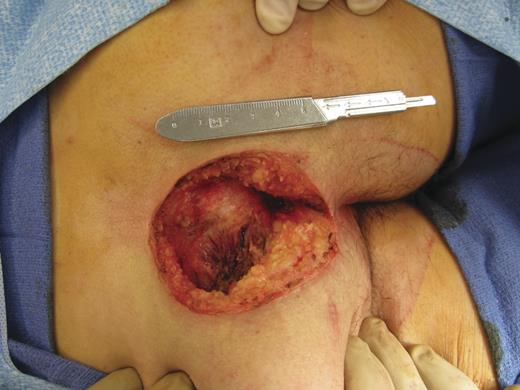
Nine consecutive cases over 2011 and 2012 of complex open pilonidal wounds were treated with MatriStem xenograft extracellular matrix material at the time of surgery and in subsequent office visits. Each of the cases involved a large, wide excision of the pilonidal disease. The average volume of the open wound after wide excision measured 9 cm in length, 7 cm in width and 4 cm in depth down to the sacrococcygeal periosteum. Each of the wounds was initially treated with MatriStem xenograft material using the eight-layer MatriStem grafts sutured to the base of the wound and the addition of 1000 mg of MatriStem powder-ground extracellular matrix material. All procedures were performed in the prone position under general anesthesia. After placement of the xenograft material, a moistened Vaseline gauze, Xeroform dressing and aqueous gel for hydration was applied, followed by gauze dry dressings for absorption of secretions. Each patient was instructed to leave the dressing intact and avoid immersion in water.
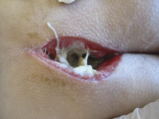
Each patient was then seen in the office on a weekly basis until the wounds were completely healed. At each visit, the packing was removed and the wound was inspected. At each of the initial visits, sufficient xenograft material remained in place and no further graft material was added. In subsequent weeks, additional MatriStem xenograft material was added in the form of 500 mg of MatriStem powder and single layer wound sheets. In each case when new graft material was added, hydrating gel and Xeroform moistened gauze was placed over the graft followed by dry, absorbent gauze. After each in-office dressing change, the patient was again instructed to avoid full immersion under water, but was encouraged to resume daily activities. Photographs were taken at each weekly visit.
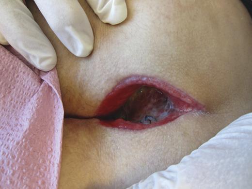
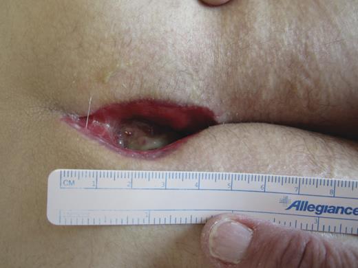
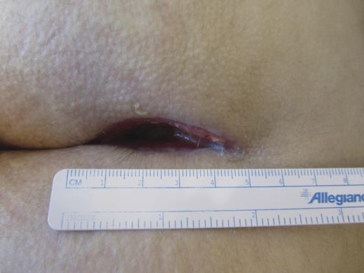
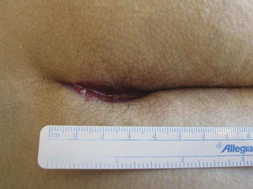
Of the nine patients, eight were male with a mean age of 23 years. Each of the nine wounds healed completely over the course of the next 5–10 weeks following wide excision of the pilonidal disease. The patients did not perform any dressing changes at home nor require any specific home wound care. The patients did not require any additional apparatus or equipment. The dressing changes were performed in the office without any sedation or anesthesia, and the patients tolerated the dressing changes well. Each of the wounds healed completely over the ensuing 5–10 weeks with this method. The average total duration of time to complete healing was 7.4 weeks. Figures 1–6 represent the progression of wound healing of Patient 7 representative of the series. Patients were followed up clinically for a median time of 12 weeks.
DISCUSSION
In this case series, we report nine cases of complete healing of complex open pilonidal sacrococcygeal wounds with MatriStem porcine-derived extracellular matrix. The average time to healing, 7.4 weeks, compares favorably with historical controls using the most common methods of dressing changes or vacuum-assisted devices. In this small series, no patients required re-operation and none developed infection. Additionally, no patients required home wound care, home wound dressing changes or additional equipment or accoutrements. The patients were able to resume their home daily activities and return for weekly wound care visits in our office. We believe that the treatment of open complex pilonidal wounds with the MatriStem xenograft material offers a compelling potential advantage compared with conventional treatments of these large wounds. The time to healing appears to be favorable when compared with historical controls. The process is a quite simple one that does not involve additional surgery or flaps. There is a significant advantage in avoiding dressing changes at home or daily visits to a wound clinic for dressing changes. Alternative techniques such as dressing wound care with daily gauze dressing changes typically requires 6 months for complete wound healing and involves a significant rate of surgical site infections and return to surgery. Alternative therapies involving flap surgery have reported to demonstrate good success with a shorter duration of wound healing [6], averaging 18 days in one study, but with a significant 23% rate of impaired wound healing and 13% wound infection rate [3, 6, 9, 10]. Strategies involving a wound vac are expensive, cumbersome for the patient and are more challenging because of the sacrococcygeal anatomic location near the anus and difficulties achieving a vacuum seal. Larger cohorts and longer term follow-ups will be needed in the future to verify these observations.
Conflict of interest Statement
The authors have no relationship with a commercial company nor have direct financial interest in the subject matter or materials discussed in this article.


