-
PDF
- Split View
-
Views
-
Cite
Cite
Tennika M. Jacobs, Andreas L. Lambrianides, Inflammatory fibroid polyp presenting as intussusception, Journal of Surgical Case Reports, Volume 2013, Issue 2, February 2013, rjt005, https://doi.org/10.1093/jscr/rjt005
Close - Share Icon Share
Abstract
The majority of adult intussusceptions have a well-defined pathological abnormality as the lead point. We present the case of a 41-year-old female who presented to the Emergency Department on four different occasions with intermittent epigastric pain, associated with vomiting. On the fourth occasion, she was found to have a bowel obstruction caused by an ileocolic intussusception, diagnosed on CT. The lead point for her intussusception was a rare non-neoplastic submucosal lesion seldom found in the ileum, an inflammatory fibroid polyp.
INTRODUCTION
In adults, intussusception causes only 1% of all bowel obstructions [1]. In the small bowel, benign lesions account for ∼70%, with malignant lesions (primary or metastatic) responsible for the remainder [2]. In contrast, intussusception in the large bowel is more likely to have a malignant aetiology, representing 66% of cases [2].
We present the case of a 41-year-old female presenting with sub-acute symptoms preceding an intestinal obstruction caused by an ileocolic intussusception with an inflammatory fibroid polyp acting as the lead point.
CASE REPORT
A 41-year-old lady presented to the Emergency Department on four different occasions with a 3-month history of intermittent epigastric pain. There was no relevant past medical history, no family history of colorectal carcinoma and her only previous surgery was a laparoscopic appendectomy 3 months earlier for acute appendicitis.
On her third presentation to the Emergency Department, she underwent an abdominal ultrasound, which displayed gross thickening of presumed large bowel (caecum/ascending colon) 48 mm in diameter and 114 mm in length. On examination, a palpable mass in the right lower quadrant was noted. However, her symptoms resolved and she was discharged home without further investigation.
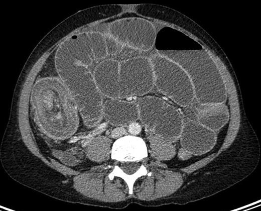
She represented one week later with severe abdominal pain associated with vomiting, poor oral intake and not opening her bowels or passing flatus for one week. On this presentation, a small bowel obstruction with multiple air fluid levels was evident on abdominal x-ray. A CT abdomen was performed, which demonstrated a target-shaped soft tissue mass pathognomonic of intussusception (as shown in Fig. 1).

CT image showing the ‘multiple concentric rings’ sign with the central cylinder representing the canal and wall of the intussusceptum; the middle cylinder representing the mesenteric fat and the other cylinder the returning intussusceptum and the intussuscipiens.
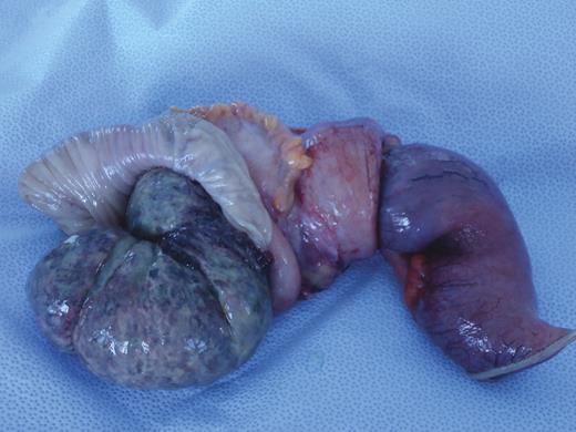
A subsequent laparotomy through a midline incision showed clear free peritoneal fluid and an ileocolic intussusception (as shown in Fig. 2).

Excised specimen, opened to illustrate ileocolic intussusception.
She underwent a right hemi-colectomy with a side-to-side ileocolic anastomosis. She received Total Parental Nutrition post-operatively for malnutrition and was discharged home following an uneventful post-operative recovery.
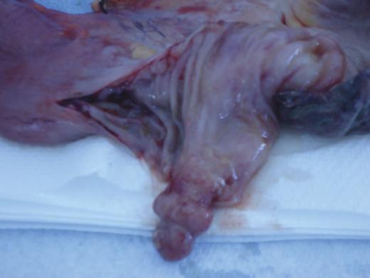
Macroscopic examination revealed a polypoid lesion measuring 25 by 10 mm, located within the terminal ileum 180 mm from the ileocaecal valve (as shown in Fig. 3).

Macroscopic submucosal polypoid lesion at the lead point of intussusception.
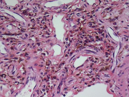
The polyp was confined to the bowel submucosa with no extension into the muscularis propria. Associated with the lesion there was localized mild predominantly chronic inflammatory cell infiltrate. Immunohistochemistry performed on this lesion was negative for S100, desmin and CD 117, effectively excluding a neurogenic tumour, gastrointestinal stromal tumour and a submucosal leiomyoma. Spindled cells within the submucosa expressed CD34 (weak to intermediate expression). This case lacks the myxoid stroma and perivascular concentric arrangement of inflammatory cells, which is best described in the stomach (as shown in Fig. 4). These features favour a terminal ileal inflammatory fibroid polyp, which was responsible for the intussusception and subsequent small bowel obstruction.

Depicting predominantly submucosa with a proliferation of bland spindle cells together with variably sized vascular channels and a scattered mixed chronic inflammatory cell infiltrate.
DISCUSSION
Adult intussusception is caused by a well-definable pathological abnormality in 70–90% of cases [2]. In general, benign lesions are more commonly the cause in the small bowel, accounting for ∼70% [2]. Examples of benign lesions include submucosal lipomas, Peutz Jeghers polyps, congenital band adhesions, intussuscepting Meckel diverticulum and inflammatory fibroid polyps [2].
Inflammatory fibroid polyps are rare benign lesions, first described by Vanek in 1949 as a gastric submucosal granuloma with eosinophilic infiltration [3]. Confusion in the literature stems from the number of different names this rare benign lesion is known by, including Vanek's tumour, eosinophilic granuloma, fibroma with eosinophlic infiltration, haemangiopericytoma and polypoid myoendothelioma [4]. Helwig and Ranier devised the generally accepted term, inflammatory fibroid polyp in 1953 [5].
The most common site for inflammatory fibroid polyps is the gastric mucosa, accounting for ∼70% [4]. Of other gastrointestinal sites affected, the small bowel is the most common, accounting for 23%, with the ileum predominating [4]. The colon (4%), gallbladder, oesophagus, duodenum and appendix have also been described as rare sites [4]. The fifth to seventh decade of life is the most common age at which patients present, both sexes being equally affected [4]. Most lesions are <2 cm in diameter, multiple synchronous or metachronous lesions being rare [4].
The aetiology of inflammatory fibroid polyps is unknown. Triggers including a foreign body, parasite and chronic H. pylori infection have been suggested but remain unsupported and theoretical [4]. A poorly controlled inflammatory response to a chemical, traumatic or metabolic mucosal injury has also been hypothesized [6]. Given its marked eosinophilic infiltration in most cases, a localized variant of eosinophilic gastroenteritis is another proposed aetiology [7]. The current consensus is a benign reactive occurrence in response to an unknown irritant [4].
Histologically, an inflammatory fibroid polyp is distinguished by a localized proliferation of mononuclear spindle-shaped cells with an inflammatory infiltrate [4]. Immunohistochemical staining shows positivity for CD34, suggesting that these polyps may develop from primitive perivascular or vascular cells [7]. Negative staining to S100 protein and CD 117 (KIT gene product) distinguishes an inflammatory fibroid polyp from a neurogenic tumour or gastrointestinal stromal tumour (GIST) [4].
The clinical presentation of an inflammatory fibroid polyp depends on the site of the tumour. Many are asymptomatic and are identified incidentally during endoscopy or laparotomy [6]. Inflammatory fibroid polyps in the small bowel can present with chronic episodes of colicky abdominal pain, lower gastrointestinal bleeding, anaemia and rarely bowel obstruction due to intestinal intussusception [7].
A discussion highlighting the histological and immunohistochemical features of inflammatory fibroid polyps, which acted as the lead point for this case of intussusception, aims to make other surgeons aware of this uncommon entity.
Acknowledgements
The authors thank Dr Tanya Wood (FRANZCR) from the Redcliffe Imaging Department for her descriptions of the ultrasound and CT images within this report. They also thank Dr David Godbolt from The Prince Charles Hospital Pathology Department for his histology images and review of the article.


