-
PDF
- Split View
-
Views
-
Cite
Cite
M Upadhyaya, E Cusick, Bilateral inflamed paratubal cysts, Journal of Surgical Case Reports, Volume 2012, Issue 9, September 2012, Page 12, https://doi.org/10.1093/jscr/2012.9.12
Close - Share Icon Share
Abstract
Paramesonephric duct remnants are an infrequent cause of abdominal symptoms in childhood. Preoperative diagnosis is often difficult and diagnosis is usually made at surgery. We report a rare presentation of an acute abdomen in a child with bilateral inflamed fimbrial cysts. Ultrasound revealed the presence of a multicystic lesion behind bladder. It was only at laparotomy the diagnosis of bilateral inflamed fimbrial cysts was establsihed. These were excised and the child made an uneventful post operative recovery.
INRODUCTION
Cysts related to the Mullerian system usually occur in paraovarian (1) or paratubal regions (2). They rarely become symptomatic in childhood (3). The symptoms are usually due to their size or torsion (4,5,6).
We are describing a case of bilateral inflamed paratubal cysts. To our knowledge this has not been described in the English literature.
CASE REPORT
An eleven year old girl presented to the emergency department with a twenty four history of abdominal pain, fever and nausea. She had also complained of loose stools and difficulty in micturition. She had been catheterised at her local hospital prior to transfer. This was the first episode of such pain. She had not attained menarche.
Her previous medical history was complex. She was born at thirty one weeks gestation. She had several laparotomies and stoma formation for necrotising enterocolitis. The stoma was finally closed at two years of age. She was born with Tetralogy of Fallot which was operated upon. Her other problems included bilateral occipital periventricular leukomalacia with visual impairment. She also had hydrocephalus and required ventriculoperitoneal shunt insertion which was complicated by meningitis. The shunt was subsequently removed. She had a normal karyotype (46XX) with no chromosomal abnormality.
On presentation she was clinically dehydrated, pyrexial, and the lower abdomen was full with tenderness in the right iliac fossa and hypogastrium. Inflammatory markers showed elevated C-Reactive Protein of 331 mg/ml (normal <10 mg/ml) and a total white cell count of 29.7 x 10⁹/L ( range 5- 13.0) with a neutrophilia of 27.0 x 10⁹/L( range: 1.5- 8.0). She had an indwelling catheter and urine dipstick was normal.
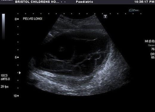
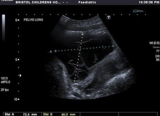
USS showed a septated cystic structure behind the bladder and abutting the ovary. There was echogenic fluid within the cysts. (Fig 1&2)


Following resuscitation she underwent laparotomy. Intraoperatively there were dense adhesions with no discernible peritoneal cavity. A large multiloculated cyst was identified at the fimbrial end of each fallopian tube. The right one measured 10x8x8 cm and the left one measured 8x6x6 cm. The fallopian tubes were dilated, inflamed and thickened. Fluid within the cysts was purulent. The cysts were excised, preserving the fallopian tubes.
Her post operative recovery was uneventful. Fluid from the cyst contained neutrophils and degenerate macrophages. On gram stain only a few white blood cells were seen and fluid culture did not identify an organism.
The histopathological analysis revealed both cysts to be lined focally by ciliated and non-ciliated epithelium forming plicae. However, for the major part, the cysts were lined by acute inflammatory exudate. The wall was composed of a thin layer of smooth muscle. The features were suggestive of inflamed Mullerian cysts.
DISCUSSION
In the paratubal and paraovarian regions the majority of the cysts are of Mullerian origin and can account for up to 76% of all the cysts (3). They are more common in adults and only occur rarely in adolescents (4%) (3). Dilatation of these cysts could be anticipated after menarche, due to the secretory activity of the lining epithelium under the influence of the hormones (3).
Large cysts could cause symptoms such as nausea, pain and vomiting (4). Some undergo torsion (5,6) with severe pain and other may also cause torsion of the fallopian tubes (2). In large adult series pelvic pain and menstrual irregularities also occurred in 57% of patients (6).
A neoplastic potential has been identified (6,7) with the incidence varying from 1.69% to 2 %. Hence excision should be seriously considered (7).
No case of inflamed paratubal cysts appears in the English literature. We can only postulate that contributory factors in our case could have included low grade infection due to previous meningitis and VP shunts.
Accurate preoperative diagnosis can be difficult. Ultrasound has been used as the first line for diagnosis. It shows a wide range of findings (8), but the majority are simple cysts. Multilocular cysts, as in our case are very rare accounting for only 4% of the cases (8). Occasionally a normal ovary abutting a cyst gives a clue to the origin of these cysts but accurate pre op diagnosis remains rare.
MRI may give more detailed three dimensional information but essentially features are the same as an ultrasound. Kishimota et al suggested that most paraovarian cysts were homogenous, near the ipsilateral round ligament and uterus (9).
Laparoscopy has been used successfully in paediatric practice to deal with large or complicated paraovarian cysts (4). The indication for open surgery is malignancy or suspicion of malignancy, dense adhesions or large cysts (6), as was the case in our patient. In our patient laparoscopic surgery would have been impossible due to the obliteration of the peritoneal cavity.
In summary we report a very rare case of bilateral inflamed fimbrial cysts which were identified and excised during laparotomy for an acute abdomen.


