-
PDF
- Split View
-
Views
-
Cite
Cite
L Meecham, V Koo, PH Rajjayabun, Uretero-iliac artery aneurysm fistula: A rare but fatal cause of haematuria, Journal of Surgical Case Reports, Volume 2012, Issue 8, August 2012, Page 16, https://doi.org/10.1093/jscr/2012.8.16
Close - Share Icon Share
Abstract
Visible haematuria from an uretero-iliac artery aneurysm fistula (UIAF) offers a diagnostic challenge and early accurate diagnosis can have a significant impact on prognosis. We report a 90 year old gentleman who presented with visible haematuria and clot retention. He required catheterisation, bladder washout and a blood transfusion. Subsequent imaging revealed an abdominal aortic aneurysm and bilateral iliac artery aneurysms with left sided hydronephrosis and hydroureter. There was no radiological evidence of a fistula between the left ureter and iliac aneurysm. Due to associated co-morbidity surgical intervention was not deemed suitable. Although his haematuria initially settled he developed catastrophic haematuria and died. Post-mortem confirmed UIAF causing fatal haemorrhage. In this report we discuss the diagnostic challenges and management options for this condition.
INTRODUCTION
Macroscopic or visible haematuria is a common urological presentation with several aetiologies including malignancy, urolithiasis, infection and trauma. The majority of causes for acute haematuria are seldom life-threatening, however, rarer diagnoses can be potentially fatal. We report a case of uretero-iliac aneurysm fistula (UIAF) causing an acute fatal haemorrhage.
CASE REPORT
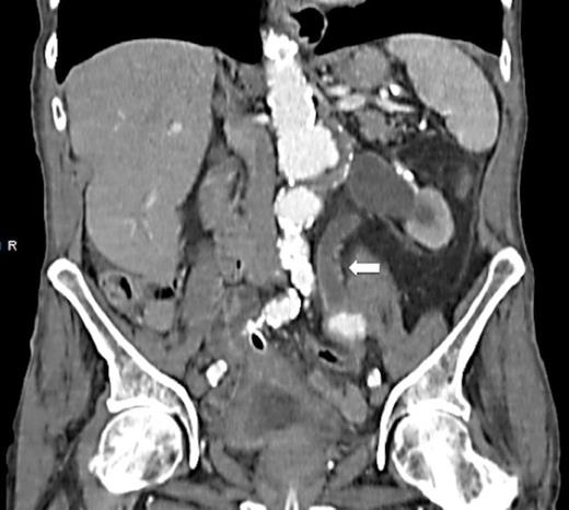
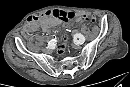
A 90 year old gentleman was admitted with haematuria and acute painful urinary clot retention. Prior to admission, he reported a 6 month history of intermittent visible haematuria. His past medical history included Duke’s A rectal carcinoma, hypertension and ischaemic heart disease. His haemoglobin count was 8.7g/dL and his renal function was normal. He was catheterised using a 3-way catheter with supplementary bladder washouts and received a 2 unit blood transfusion. Abdominal ultrasound revealed moderate left hydronephrosis. A CT urogram detected a 48mm infra-renal aortic aneurysm with bilateral iliac artery aneurysms – 33mm left, 31mm right. This scan also identified moderate to severe hydronephrosis on the left side with hydroureter extending down to the level of the iliac aneurysm (Fig. 1 & 2). There was no obvious fistula between the left ureter and the iliac aneurysm.

CT scan demonstrating hydronephrosis and hydroureter on the left side
The patient was referred to the vascular surgical team for consideration of repair of the left iliac aneurysm. In view of his poor general health and co-morbidities he was deemed unsuitable for surgical intervention. Ureteric stent insertion was not necessary as he was asymptomatic with normal renal function. After a 5 day period his haematuria resolved and a subsequent flexible cystoscopy was generally unremarkable.

CT scan demonstrating hydronephrosis and hydroureter on the left side
After 14 days the patient was fit for discharge, however, he suddenly collapsed after developing further heavy visible haematuria. There was a clinical suspicion of iliac aneurysm rupture with an evolving fistula into the left ureter. He was reviewed by the vascular team who concluded that surgical intervention was futile. He was kept comfortable and died shortly afterwards. A formal hospital post-mortem confirmed an UIAF on the left side. There was significant blood in the bladder but none in the abdomen. The cause of death was exsanguination through an UIAF.
DISCUSSION
Iliac artery aneurysms are usually asymptomatic and can be incidentally found coexisting with abdominal aortic aneurysms. They can remain undetected until pressure is exerted upon local surrounding structures. In this case, the iliac artery aneurysm caused extrinsic compression of the left ureter resulting in hydronephrosis. After a period of time an ensuing cascade of inflammatory events may lead to the development of a fistula between the aneurysm and ureter. It is postulated that initially the integrity of the vasa vasorum is disrupted with weakening of the tunica adventitia and media. This increases the susceptibility of the aneurysm to necrosis and rupture (1). Additional contributory factors hastening the development of fistulae include pelvic radiation, direct trauma from pelvic surgery, ureteric instrumentation and ureteric stenting (2,3)
Although a rare entity, the incidence of UIAF is increasing. This is thought to be related to modern regimes of aggressive surgery and radiation in the management of pelvic malignancy and frequent use of ureteric stents (4). Investigations for an UIAF include CT angiography, MR angiography, selective angiography, diagnostic ureteroscopy and retrograde ureteropyelography. Despite these imaging modalities it can be challenging to formally identify a fistula as these can be small and may not be revealed at the time of imaging. A high index of clinical suspicion for a UIAF is paramount to the diagnosis and can have a significant impact on prognosis. Previous studies have demonstrated a high mortality rate (64%) in patients who underwent exploratory laparotomy without adequate preoperative diagnosis. This was reduced to 0% in patients where the diagnosis was considered prior elective surgery (5).
A multidisciplinary team approach involving urologists, vascular surgeons and radiologists is important in the treatment of uretero-iliac aneurysm fistula (3). Management involves concurrent repair of arterial and ureteric aspects of the fistula. The arterial repair can be carried out by primary open repair, arterial ligation or selective vascular embolization with extra-anatomical vascular management. Where renal function is adequate options include ureteric resection with primary anastomosis and nephrostomy or stent insertion. In a poorly functioning kidney nephro-ureterectomy may be considered.
Although a rare entity, UIAF remains a diagnostic challenge. A high index of clinical suspicion and accurate diagnosis may avert a potentially catastrophic outcome.


