-
PDF
- Split View
-
Views
-
Cite
Cite
M Al-Omran, Repair of a true dorsalis pedis artery saccular aneurysm, Journal of Surgical Case Reports, Volume 2012, Issue 7, July 2012, Page 15, https://doi.org/10.1093/jscr/2012.7.15
Close - Share Icon Share
Abstract
Aneurysms of the foot arteries are rare. A case of a true dorsalis pedis artery saccular aneurysm in a 60 years old man in which there was no history of trauma is presented. The arterial duplex, intraoperative features, operative procedure and literature review are presented and discussed.
INTRODUCTION
Dorsalis pedis artery (DPA) aneurysms, though uncommon, are well recognized clinical presentation and it was first described by Cuff in 1907 (1). Then after, many case reports have described this rare aneurysm and proposed different management modalities.
This article presents a case of true DPA saccular aneurysm in a 60 year old man and discusses and summarizes the different clinical presentations, proposed aetiology, investigations modalities, and treatment options in this rare condition compared to the previously reported cases.
CASE REPORT
A 60 years old man presented with a painless pulsatile mass located on the dorsal part of the right foot. The mass was incidentally discovered by his family physician during a routine follow-up 3 years prior to the referral. Over that time, the mass had enlarged, but there were no symptoms related to the mass. There was no history of blunt or penetrating trauma, nor surgery in the foot. The patient is known to have diabetes mellitus on oral hypoglycemic medication, hypertension, and mild renal function impairment. Family history of aneurysmal disease was negative.
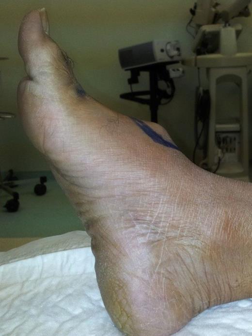
On physical examination, the patient appeared healthy. Right foot examination showed pulsatile, non-tender and compressible mass on the dorsum of the foot (Fig. 1). There was no thrill or bruit over the mass. Posterior tibial artery was palpable at ankle. No signs of toes ischemia were observed. No other vascular abnormalities were detected elsewhere.

Dorsum of the right foot showing a pulsatile mass suggestive of a dorsalis pedis artery aneurysm
Arterial duplex study was performed for the abdominal aorta and bilateral lower limbs which revealed no aneurysms in the aorta nor the popliteal arteries, and confirmed the presence of a right DPA aneurysm. The aneurysm was 2.6 cm in diameter and 2 cm in length with a small mural thrombus. Right foot toes pressure measurement showed normal readings.

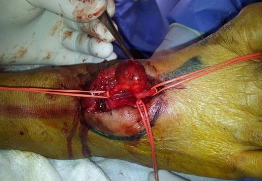
Intra-operative Image: Saccular aneurysm of the right dorsalis pedis artery
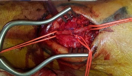
Surgical exploration was carried out under ankle block. A longitudinal skin incision was made directly over the aneurysm. Sharp dissection through fascia revealed a 3 cm DPA saccular aneurysm and it was controlled proximally and distally using vessel loops (Fig. 2). A trial of intraoperative clamping of the aneurysm proximally and distally resulted in decreased Doppler signals in digital arteries. Therefore, the aneurysm was resected and an end-to-end anastomsis was done (Fig. 3). A good pulse was felt and Duplex scan confirmed the artery to be patent during immediate post-operative period. A posterior slap was applied to immobilize the ankle for 1 week.

Intra-operative Image: Post resection of the aneurysm and reconstruction by end-to-end anastomosis
The postoperative course was uneventful, and the patient was discharged home on the second postoperative day. Histology demonstrated a true aneurysm where all three layers (intima, media, and adventitia) of the arterial wall were seen and showed focal degenerative changes. Nine months postoperatively, the patient was seen in the clinic with normal right foot function and good palpable pulse at DPA and good perfusion as demonstrated by arterial duplex study and toes pressure.
DISCUSSION
Dorsalis pedis artery (DPA) aneurysm, though uncommon, has been classically reported in the literature as either true aneurysm or psudoaneurysm secondary to trauma. A computerized literature search was conducted in MEDLINE (up to March 2012). The search used the keywords dorsalis pedis artery and aneurysm with no limits to articles date of publication, type, language, gender nor age group (2). Thirty one cases of DPA aneurysm have been reported. Of these, 18 cases (58%) were reported as psudoaneurysm of DPA secondary to trauma or iatrogenic causes such as orthopedic surgery and cannulation of the artery.
Patients with DPA aneurysm are usually presents with painless pulsatile mass in the dorsum of the foot, however more serious presentations such as rupture aneurysm or acute ischemia of the forefoot have been reported (3,4).
The diagnostic and treatment algorithms for DPA aneurysm are not well established, because the disease is rare. The use of vascular laboratory tests such as Doppler ultrasound and arterial duplex poses many advantages as had been presented. First, the tests are not invasive, less expensive and do not require the use of contrast which is a very important in our patient who has mild renal impairment. Second, they can delineate and localize the DPA aneurysm with accuracy and they can identify the presence of mural thrombus. Finally, they are of great help in the assessment of the adequacy of foot perfusion perioperatively, and therefore, help in the decision of whether to ligate or to reconstruct the aneurysm. Although the use of magnetic resonance imaging (MRI) has been advocated in the diagnostic workup for foot arteries aneurysms (5), it has some disadvantages that include the low quality in visualizing digital arteries. Conventional arteriogram, on the other hand, has the ability to visualize small arteries and can aid in the planning of surgery especially with selective posterior tibial and dorsalis pedis arteries angiogram. In addition, it can be used as a therapeutic option with the use of embolization of favorable lesions.
Treatment of DPA aneurysms should not be based on the presence of symptoms, because the development of thromembolic complications with subsequent toes and forefoot ischemia can occur without warning signs or even the risk of rupture (3,4). The surgical options for DPA aneurysms depend on the presence of adequate perfusion in the forefoot after excluding the aneurysm from the foot circulation. If the forefoot and toes are adequately perfused and the posterior tibial artery is intact, simple resection is the surgical option. However, if the foot perfusion is inadequate, DPA reconstruction is mandatory. In the present case, the DPA aneurysm was resected with reconstruction because of the intra-operative evidence of inadequate toes perfusion after excluding the aneurysm from the foot circulation as documented by poor Doppler signals in all digital arteries.


