-
PDF
- Split View
-
Views
-
Cite
Cite
Svetlana Kleyman, Lelia Logue, Vincent Lau, Erick Maio, Aliu Sanni, Feroze Khan, Ileal diverticulitis: an uncommon diagnosis for right lower quadrant pain, Journal of Surgical Case Reports, Volume 2012, Issue 11, November 2012, rjs010, https://doi.org/10.1093/jscr/rjs010
Close - Share Icon Share
Abstract
We report an interesting case of ileal diverticulitis which posed a diagnostic challenge. A 75-year-old female presented to the emergency department with severe right lower quadrant pain for 3 days. The clinical history, examination and imaging suggested a diagnosis of acute appendicitis. The patient was taken to the operating room for an open appendectomy. The intra-operative findings demonstrated a large mass at the ileocecal junction involving the appendix as well as multiple nodular masses in the ileum and cecum. The patient underwent a right hemicolectomy with ileocecal anastomosis. The pathology result revealed Ileal diverticulitis. Ileal diverticulitis is a rare form of diverticulitis. It can often mimic other processes such as acute appendicitis. Once ileal diverticulitis is diagnosed, it should be treated with the same principles as for sigmoid diverticulitis. Though rare, ileal diverticulitis should be considered in the differential diagnosis of a patient who presents with right lower quadrant pain, and a computed tomography scan that shows an inflammatory process in the right lower quadrant, in the setting of a normal appendix.
INTRODUCTION
Ileal diverticulitis is a diagnosis that is rarely encountered. These diverticula are false diverticula. Most patients with ileal diveritculosis are asymptomatic. When ileal diverticulosis becomes diverticulitis, the patient may present with right lower quadrant pain and a clinical picture that leads to suspicion of acute appendicitis. If ileal diverticulitis is diagnosed on computed tomography (CT) scan, the patient should be treated in a nonoperative manner with intravenous antibiotics and bowel rest.
CASE REPORT
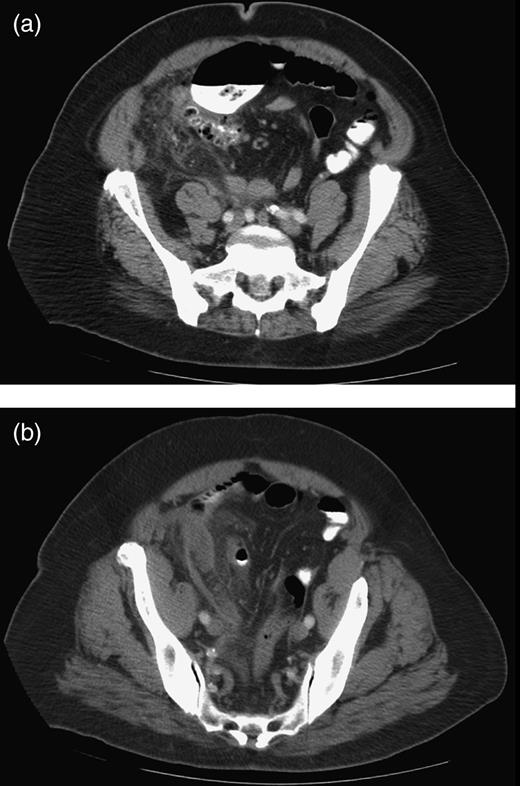
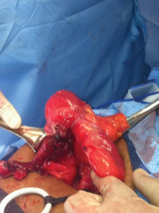
A 75-year-old obese African-American woman presented to the emergency department with progressively worsening, severe and constant aching right lower quadrant pain for 3 days. She described the pain as starting in the epigastrium and moving to the right lower quadrant after 1 day. She vomited once 2 days ago, but denied current nausea, vomiting or diarrhea. Her last bowel movement was 1 day prior to admission. She denied fever, chills and urinary symptoms. Her past medical history included hypertension, diabetes mellitus and hypercholesterolemia. On examination, she was afebrile with stable vital signs. Palpation of the right lower quadrant elicited rebound tenderness and guarding. Bowel sounds were normoactive. Laboratory investigations were essentially within the normal range. Abdominal computed tomographic scan with oral and intravenous contrast media showed an inflammatory mass in the right lower quadrant and a small amount of free fluid on the right side of the pelvis and in the cul-de-sac (Fig. 1a and b). The appendix was not specifically identified. The patient was given a dose of Cefotetan and taken to the operating room for an open appendectomy. A transverse incision was made at McBurney's point and the appendix was visualized. It was noted that the appendix was extremely inflamed and thickened with mesentery down to the cecal base. Further exploration showed a large mass at the ileocecal junction involving the appendix (Fig. 2). The appendix also had a necrotic base. In addition, multiple nodular masses were observed in the ileum and cecum. Because a carcinoma could not be excluded, the decision was made to perform a right hemicolectomy with primary anastomosis. Pathology revealed Ileal diverticulitis.

(a and b) Axial CT scan demonstrating infiltrative changes in the pericolonic fat and a small amount of free fluid in the right pelvis.

Intraoperative findings demonstrating a large mass at the ileocecal junction involving the appendix.
DISCUSSION
Ileal diverticulitis is an uncommon entity, occurring with an incidence of 0.1–1.5% [1].
Ileal diverticula are false diverticula when compared with the more common true Meckel's diverticulum occurring in the ileum. They are usually multiple and occur at the mesenteric border, sometimes hidden in the mesentery and overlooked during surgery [2]. Diverticulosis is a condition of patients after their sixth decade of life [3]. Acquired diverticula of the ileum may be a primary condition or secondary due to abdominal surgery, tuberculosis or Crohn's disease. Most patients are asymptomatic and diagnosis is made on routine imaging studies or at autopsy. Of the patients who become symptomatic, most present with intermittent mild abdominal discomfort that may localize to the right lower quadrant. Complications include diverticulitis, intestinal obstruction, volvulus, fistula, bleeding or perforation. Because of the similar presentation, ileal diverticulitis can mimic appendicitis. This results in many cases going to the operating room without an accurate preoperative diagnosis. The acute complications of ileal diverticula are very rare, occurring in only 6.5–10.4% of the cases. The mortality of the associated complications is 25–50% [4]. Ileal diverticulitis is diagnosed with radiological studies. CT is the initial test of choice demonstrating wall thickening, extraluminal free air, mesenteric inflammation and fluid collection if perforation has occurred. In the presented case, the CT showed an inflammatory mass and a small amount of free fluid on the right side of the pelvis and in the cul-de-sac. There was no free air and no mention of ileal diverticula. The appendix was not well visualized, but a diagnosis of suspected appendicitis was made as is the mistake commonly made with ileal diverticulitis because of the similar presentation. Once a diagnosis of ileal diverticulitis is made on CT, an enteroclysis should be performed to evaluate for other small bowel diseases such as Crohn's. Plain upright X-rays can also play a role early on in the case. It may demonstrate a pneumoperitoneum suggestive of a gastrointestinal perforation.
The management of ileal diverticulitis is similar to that of colonic diverticulitis. It should be treated conservatively whenever possible with bowel rest, IV hydration and IV antibiotics. For those who present with complications such as bleeding, obstruction or perforation, surgical intervention is required. The procedure involves segmental resection with primary anastomosis [1]. In conclusion, ileal diverticulitis, although rare, should be included as a differential in all cases of right lower quadrant pain. The index of suspicion should be high when CT findings show an inflammatory process but not suggestive of appendicitis. Simple cases should be managed conservatively, but complicated cases require immediate surgical intervention in order to decrease morbidity and mortality.


