


Abstract
A congenitally absent unilateral submandibular gland is a rare condition. We present such a case with an associated ipsilateral benign hyperplastic sublingual gland. The enlarged sublingual gland may well represent a compensatory response to the missing submandibular gland, but it is known that sublingual gland tumours are malignant in approximately 90% of cases and so a sublingual gland swelling is viewed with a high degree of suspicion. Clinical diagnosis of sublingual hyperplasia becomes a challenge if there is a missing unilateral submandibular gland, but a full investigation is important to rule out a more sinister pathology.
INTRODUCTION
Absent major salivary glands are rare occurrences; Iguchi and Uyama (1) reported a case of bilateral submandibular gland aplasia, this was associated with a unilateral submandibular haemangioma. Ahmed M and Strauss M et al (2) also reported bilateral submandibular gland aplasia with associated prolapsing sublingual salivary tissue through mylohyoid muscle.
CASE PRESENTATION
A 57-year old male, non-smoker with no significant medical history was referred by the community dentist to the maxillofacial department with an asymptomatic left floor of mouth swelling, which was noticed by the practice hygienist.
Intraoral examination revealed a 2cm soft and non-tender left floor of mouth submucosal swelling, in the region of the sublingual gland. Overlying mucosa was intact and with a normal appearance. There was no enlargement of the submandibular gland region and palpation of the remainder of the neck was unremarkable.
Radiographic imaging showed no evidence of opacities suggestive of calculi or any dental disease.
The clinical impression was of an enlarged sublingual gland, and so a fine needle aspiration was carried out. The cytology report confirmed salivary gland cells, some of which were enlarged. In view of this, a magnetic resonance imaging scan was ordered which confirmed a left anterior floor of mouth mass of 3 x 1 cm, (figures 1 and 2). Interestingly there was also a missing ipsilateral submandibular gland. The remaining major salivary glands were of normal appearance and dimensions.
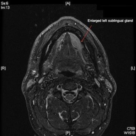
Magnetic resonance imaging scan showing enlarged left sublingual gland.
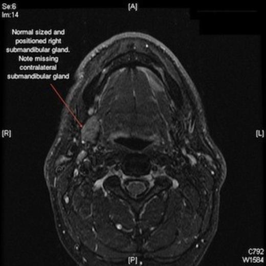
Magnetic resonance imaging scan showing missing left submandibular gland
The patient underwent removal of the enlarged sublingual gland, which dissected out with relative ease, and no other clinical pathology was noted intra-operatively.
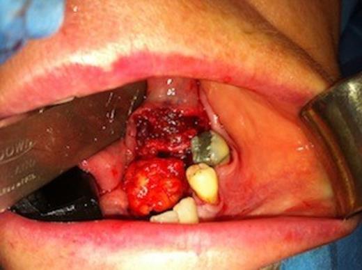
Intraoperative view of left sublingual gland
The histology report showed minor changes consistent with chronic sialoadenitis with no evidence of malignancy.
DISCUSSION
Unilateral submandibular gland agenesis is thought to be uncommon. Aiyappan et al who also reported an incidental left-sided submandibular gland aplasia in 2010 (3) conducted a literature search where they found 13 reported cases of unilateral submandibular gland aplasia from 1990 to 2009. Interestingly only one of the cases was on the left side. The actual prevalence of unilateral submandibular gland agenesis is unknown, as patients are often asymptomatic and the missing glands are often noted as incidental findings on imaging.
The cause of this unilateral anomaly is unknown; Roh et al have suggested unique defects of early fetal development (4). The hyperplasia of the ipsilateral sublingual gland may represent a compensatory response to the missing submandibular gland.
CONCLUSION
Unilateral sublingual gland hyperplasia in association with ipsilateral submandibular gland aplasia is rare. This report demonstrates compensatory enlargement of the sublingual gland, which poses a diagnostic challenge on simple clinical examination. As a large proportion of sublingual gland tumours (presenting as simple swellings) are malignant it is important to be suspicious and investigate these enlargements further to rule out a malignant pathology.
