-
PDF
- Split View
-
Views
-
Cite
Cite
Kunal Kotkar, Ravi Thakkar, MC Songra, Giant urethral calculus, Journal of Surgical Case Reports, Volume 2011, Issue 8, August 2011, Page 9, https://doi.org/10.1093/jscr/2011.8.9
Close - Share Icon Share
Abstract
Primary urethral calculus is rarely seen and is usually encountered in men with urethral stricture or diverticulum. We present a case of giant urethral calculus secondary to a urethral stricture in a man. The patient was treated with calculus extraction with end to end urethroplasty.
INTRODUCTION
Urethral calculi are already an uncommon entity and giant calculi in the urethra are extremely rare. They represent 2% of all urinary stone disease. Here we present a case of giant urethral calculus secondary to a urethral stricture in a man. The rarity of this condition prompted us to present this case.
CASE HISTORY
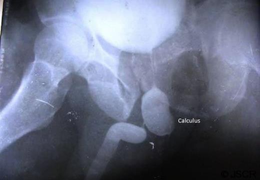
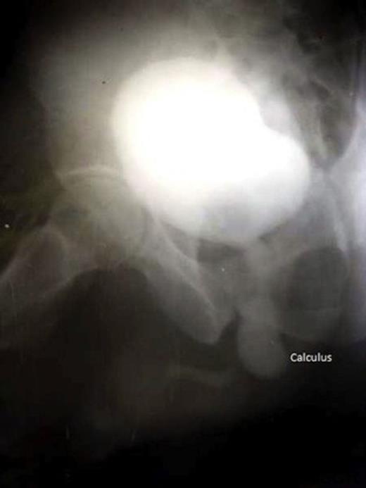
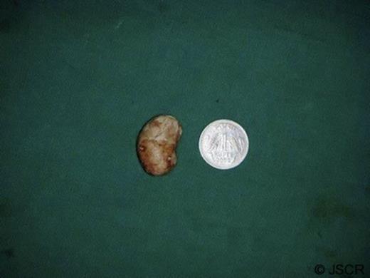
A 55 year male was admitted to our institution with complaints of pain and swelling in perineal region for 6 days and purulent discharge from the swelling for 2 days. Patient was apparently quite well 4 months prior when he noticed a progressive thinning of urinary stream along with dysuria and burning on micturition. Three months later the patient had an episode of retention of urine for which patient attended our outpatient department. A trocar suprapubic catheterisation (SPC) was performed and a 16 Fr Foley’s catheter was passed into the bladder. Patient was asymptomatic for 1 month followed by reappearance of the swelling and pain in the perineal region. On local examination there was a swelling 2 x 2 cms in size in the left perineal region with purulent discharge. Urine culture and pus culture showed growth of Klebsiella resistant to all tested antimicrobials. Patient was subjected to retrograde urethography (figure 1) and micturiting cystourethography (figure 2). Patient was diagnosed as a case of urethral calculus with urethral stricture and was posted for urethral calculus removal with urethroplasty. The calculus of 30mm x 20mm x15mm (figure 3) was extracted successfully and end to end urethroplasty was performed.


DISCUSSION
Urethral stones are classified as (a) Native or autochthonous and (b) Migrant or secondary depending upon their site of origin.(1,2)
Migrant stones are much more common and are ones which have migrated from higher up in the urinary tract.
Native stones are struvite, calcium phosphate or calcium carbonate in composition, have no nucleus and are of uniform structure. They are formed in the urethra proximal to strictures, in congenital and acquired diverticula, with chronic infection with especially urea splitting organisms or with foreign bodies. They generally do not cause acute symptoms because of their slow development. May present with a mass on the undersurface of penis, urethral discharge, dyspareunia, irritative voiding symptoms and haematuria
Migrant stones are calcium oxalate and phosphate in composition. They often cause acute symptoms causing retention, frequency, dysuria, poor stream or dribbling. Urethral calculi are preponderantly found in the prostatic urethra, the bulb, the proximal penile urethra, the fossa navicularis and external meatus.
Treatment is contingent on the size and location of calculus and condition of the urethra.
Urethroscopic lithotripsy and removal is useful in any situation.
Meatotomy may be used if stone is in fossa navicularis or external meatus
Small stones can be gently massaged with intraurethral instillation of xylocaine.
Strictures are to be dealt with, if present, with urethrotomies or urethroplasties.
Calculi in posterior urethra can be pushed back into bladder
In cases of urethral diverticulum, diverticulectomy and repair should be done.


