-
PDF
- Split View
-
Views
-
Cite
Cite
S Vaddi, VR Pogula, R Devraj, AV Sreedhar, Congenital bladder diverticulum – a rare adult presentation, Journal of Surgical Case Reports, Volume 2011, Issue 4, April 2011, Page 8, https://doi.org/10.1093/jscr/2011.4.8
Close - Share Icon Share
Abstract
Congenital bladder diverticula usually present during childhood. They are solitary and present with infection, haematuria and abdominal pain. They are associated with a smooth walled bladder without bladder outlet obstruction. An adult male presented with voiding symptoms and lower abdominal pain. On evaluation he had a grossly distended bladder extending from the hypogastrium up to the right hypochondrium. Investigations revealed a large bladder diverticulum and left hydronephrosis with a non-functioning left kidney. Left nephroureterectomy and diverticulectomy were carried out. We report this case because of its unusual adult presentation.
INTRODUCTION
Congenital bladder diverticula represent a herniation of the bladder urothelium through the muscularis propria of the bladder wall. Diverticular wall is composed of mucosa, lamina propria, scattered thin muscle fibres and an adventitial layer. Bladder diverticula generally empty poorly during micturition, leaving a large post-void residual urine volume that results in the characteristic findings on presentation and imaging.
CASE REPORT
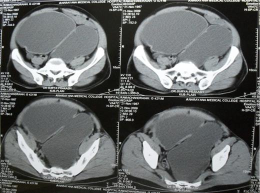
A 42 year old adult presented with poor urinary stream and lower abdominal pain over the last year. On examination he had a grossly distended bladder extending from the hypogastrium up to the right hypochondrium. External genitalia was normal. An ultrasound abdomen revealed a large bladder diverticulum in the left posterolateral aspect of bladder, mild right sided hydronephrosis with a right renal calculus and severe left hydroureteronephrosis with a thinned out cortex. Voiding cystourethrogram (VCUG) showed large bladder diverticulum with no evidence of reflux and normal posterior urethra. Intravenous urogram (IVU) and DTPA renogram revealed a non-functioning left kidney. A CT-KUB confirmed mild right sided hydronephrosis with a 1cm renal calculus, severe left sided hydroureteronephrosis and a large bladder diverticulum (Fig. 1).

Plain CT scan of abdomen and pelvis showing large bladder diverticulum posterior to the bladder
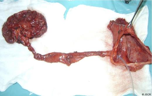
Cystoscopy showed a normal urethra, a solitary, large, wide mouthed diverticulum arising from the left posterolateral wall of the bladder and left ureteric orifice was not visualised. In view of non functioning left kidney and large bladder diverticulum with chronic urinary retention, left nephroureterectomy and diverticulectomy was carried out. Macroscopic examination of the specimen revealed that the left kidney was grossly dilated with a thinned out parenchyma and a dilated left ureter entering into the diverticulum (Fig. 2). Diverticulectomy was undertaken using a combined intravesical and extravesical approach. The patient made an uneventful recovery

Left nephroureterectomy and bladder diverticulectomy specimen with Left ureter opening into the diverticulum
DISCUSSION
Congenital diverticula usually present during childhood. (1) These are usually solitary, occur almost exclusively in boys and are located lateral and posterior to the ureteral orifice. (2) The primary causation appears to be a congenital weakness at the level of the ureterovesical junction. (3) Bladder diverticula do not contain a defined functional muscularis propria layer and therefore empty poorly with micturition..
Most bladder diverticula are diagnosed incidentally or during the investigation of nonspecific lower urinary tract symptoms, haematuria, or infection. The diagnosis of bladder diverticula relies on radiological and endoscopic findings. Voiding cystourethrography (VCUG) is an excellent study to detect bladder diverticula. (4) Upper tract imaging is to evaluate for asymptomatic or silent hydroureteronephrosis related to the diverticulum.
Symptoms or complications related to bladder diverticula are most often due to poor emptying of the diverticulum and urinary stasis. This patient presented with chronic retention due to bladder neck obstruction by the diverticulum, which was extending behind the trigone and impinging on the bladder neck. Left gross hydroureteronephrosis is probably due to left ureter opening into the diverticulum and compression on left lower ureter by the diverticulum itself. Bladder diverticulectomy is indicated for the treatment of symptoms related to the diverticulum or for the major complications directly related to it, including chronic relapsing urinary tract infection, stones within the diverticulum, carcinoma or premalignant change, and upper urinary tract deterioration as a result of obstruction or reflux. (5) Open excision is usually performed through a transvesical approach. Combined intravesical and extravesical approach is required for large diverticula.
Chronic retention of urine and non-functioning kidney secondary to a large congenital bladder diverticulum in an adult is extremely rare. Diverticulectomy restores voiding function in such cases.


