-
PDF
- Split View
-
Views
-
Cite
Cite
AJ Runia, A Schasfoort, AJH Kerver, AC van der Ham, A rare case of pyomyositis due to swine flu, Journal of Surgical Case Reports, Volume 2011, Issue 4, April 2011, Page 7, https://doi.org/10.1093/jscr/2011.4.7
Close - Share Icon Share
Abstract
Pyomyositis is a bacterial infectious disease of the large skeletal muscles, mostly seen in tropical regions. A case with such a multitude of abscesses has never been described in the western world, nor following an H1N1 infection. We report of a 31 year old man who presented himself with complaints of muscle pain and fever. His complaints were attributed to a proven H1N1 infection. However, despite proper therapy his condition worsened. He had developed multiple abscesses in both arms and legs. After surgical and radiologic drainage and antibiotic treatment our patient healed without any symptoms. Pyomositis is related with immuno-compromisation. Our patient might have been immunocompromised due to the H1N1 infection. Rhabdomyolysis following influenza has been described before, however a relation with H1N1 never was. Imaging studies help detect and confirm the diagnosis. When missed, serious complications may arise.
INTRODUCTION
Pyomyositis is charactarized by abscesses in the skeletal muscles of mainly the pelvis and lower extremities and is rarely seen in temperate regions. Due to an increase in immunocompromised patients the incidence of pyomyositis is rising in the western world. Staphylococcus aureus is the most common bacterial agent (1,2,3). We describe a case of a young man with tenderness of his muscles, caused by multiple abscesses and swine flu.
CASE REPORT
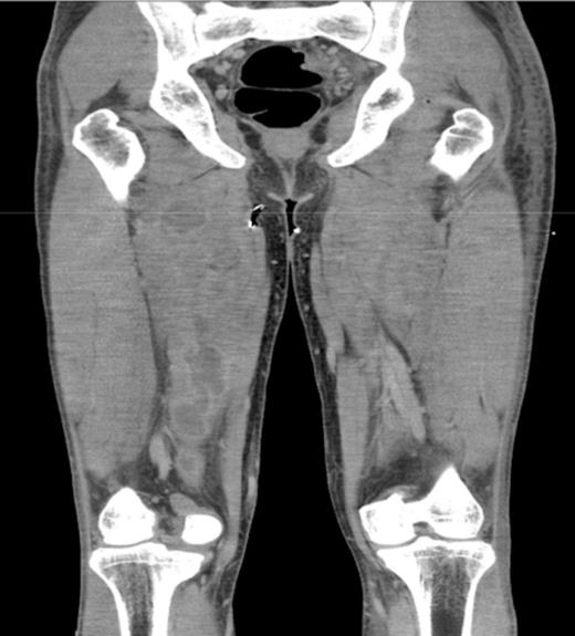
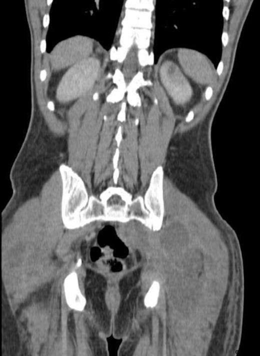
A 31 year old, otherwise healthy young man presented to our emergency department with muscle ache of his legs, a sore throat and fever. Physical examination showed subfebrile temperature, pain of both upper legs and buttocks, but no local inflammation or swelling was noticed. Laboratory examination showed slightly elevated levels of CoReactive Protein (CRP), white blood count (WBC) and Creatine Kinase (CK). The PCR for H1N1 tested positive. With the diagnosis “swine flu” the patient was admitted to the department of internal medicine and therefore oseltamivir treatment was started. However this treatment rendered no success, since the pain increased and spread to both legs and arms. Laboratory findings showed a further increase of both CRP and WBC. The subsequent CT-scan showed multiple abscesses (fig. 1, 2) in the psoas muscle, in both legs and arms. Subsequently 21 abscesses were drained, both surgically and radiologically. All wound and blood cultures showed Staphylococcus aureus, MRSA and PV negative. Our patient was also treated with intravenous flucloxacilline and rifampicine. The patient healed without destruction of muscles.


DISCUSSION
Pyomyositis was first described in 1885 by Scriba as an infectious disease of mainly the skeletal muscles (4). It can be seen in all stages of life, but most frequent in adolescents, with a male: female ratio of 3:1. Two subtypes of pyomyositis are described, a tropical and a temperate type. However, apart from geography and incidence, they do not differ. The incidence of pyomyositis in temperate regions is increasing, due to an increase of predisposing factors, such as immunocompromised patients, e.g. due to HIV, diabetes, malignancies and chronical diseases. Preceding trauma of the affected muscles, by for example fierce exercise, has also been proposed as an important predisposing factor of pyomyositis. The etiology of pyomyositis however is still unknown (2,5,6,7)
Staphylococcus aureus is cultured in 70-95 % of the cases, which often is an MRSA strand. Other microorganisms cultured are Streptococcus sp, Escherichia coli and Mycobacterium tuberculosis. Cultures and Gram-stains are therefore very important to guide proper antibiotic treatment. (1,2,3)
There are three stages of pyomyositis; stage I is characterized by vague symptoms; sore muscles, cramping, low-grade fever and discomfort. Most patients only seek medical help in stage II, because of formation of abscesses in the muscles, approximately 10-21 days after the onset of the disease. Fever, pain as well as erythema and swelling of the muscles appear. If not treated properly this will proceed into stage III, with serious illness, metastatic abscesses, sepsis, necrosis of the muscles, multiorgan failure and higher mortality. (7,8)
Diagnosing pyomyositis may be challenging, due to the vague presentation in stage I. Therefore it is often diagnosed with some delay. When the symptoms become more outspoken, apart from history and physical examination, imaging studies are usefull. CT-scan and ultrasound are used to detect abscesses. MRI is the diagnostic tool of choice, because it can detect affected muscles in an early stage. Also more abscesses can be found at unexpected sites, especially when the patient is not responding to treatment. Multifocal abscesses are rare. But, as with our patient, it is of great importance to scan the entire body to detect other abscesses in unexpected regions, if a patient does not respond adequately to the initial treatment.
Treatment of pyomyositis depends on the stage in which it is diagnosed. Stage I can be treated with adequate antibiotic therapy alone. In case of abscess formation proper surgical or radiologic drainage of all abscesses is required. The duration of administration of antibiotics is usually 7-10 days, but needs to be adjusted regarding extensiveness of the disease; number of abscesses, osteomyelitis and immune status of the patient. (1)
If treated properly, most patients heal without additional complications and low mortality rates have been described, up to 1.5%. (1)
The presentation of our patient is very extraordinary, because of the extent of the disease and the abundance of abscesses. No other case of such a multitude of abscesses have been reported. An explanation for the severity of the ilness might be found in the preceding or simultaneous “swine flu” infection. Myositis due to influenza and even rhabdomyolysis due to H1N1infection have been described (9,10). We consider muscletrauma due to the viral infection to be a predisposing factor for the extensiveness of the disease in this case. Secondly, pyomyositis is more often seen in immunocompromised patients. Swine flu is thought to negatively correlate with the immune state of the patient.
We conclude that pyomyositis can be widespread in patients, which is sometimes hard to diagnose, but should be thought off , especially in times of an influenza pandemic.


