-
PDF
- Split View
-
Views
-
Cite
Cite
A Mirza, R Gogna, M Kumaran, M Malik, AE Martin-Ucar, The surgical management of intercostal lung herniation using bioprosthesis, Journal of Surgical Case Reports, Volume 2011, Issue 2, February 2011, Page 6, https://doi.org/10.1093/jscr/2011.2.6
Close - Share Icon Share
Abstract
Lung hernia is a rare occurrence. Consequently there is little literature providing guidance to effective management. Classified as congenital or acquired, there are fewer than 300 cases described in current literature (1). We describe a unique method for the management of spontaneous rib fractures and, resulting posterior lung herniation in a 65 year old man following a bout of coughing.
INTRODUCTION
Obtrusion of lung tissue outside of the thoracic cage is an exceedingly rare entity, with fewer than 300 cases documented in current literature (1). Most herniae are diagnosed either congenitally, following trauma or post-surgery and are occasionally related to pulmonary injury. Classically, chest wall trauma has been managed with pain control alongside intensive chest physiotherapy (2). Spontaneous lung herniae are rarely described and there are few descriptions of operative management (2). Repair, particularly in the presence of a patient’s symptoms can restore parietal mechanics and is advised (4).
Obtrusion of lung tissue outside of the thoracic cage is an exceedingly rare entity, with fewer than 300 cases documented in current literature (1). Most herniae are diagnosed either congenitally, following trauma or post-surgery and are occasionally related to pulmonary injury. Classically, chest wall trauma has been managed with pain control alongside intensive chest physiotherapy (2). Spontaneous lung herniae are rarely described and there are few descriptions of operative management (2). Repair, particularly in the presence of a patient’s symptoms can restore parietal mechanics and is advised (4).
CASE REPORT
A 65 year old smoker presented with a five week history of considerable left sided pleuritic pain and shortness of breath following a severe bout of coughing. Past medical history included untreated chronic obstructive pulmonary disease, hypertension, deep vein thrombosis, bilateral total knee replacements and a laparoscopic cholecystectomy. On examination he was found to be significantly overweight and exhibited intensive bruising on his left chest with a floating portion of ribs.
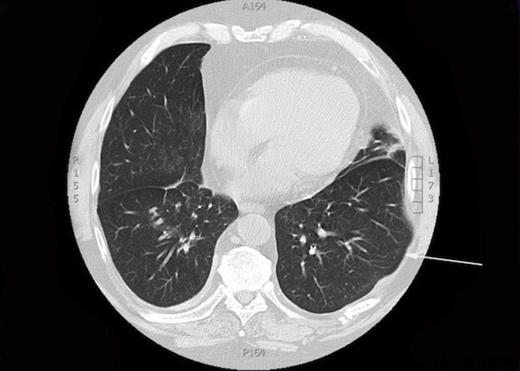
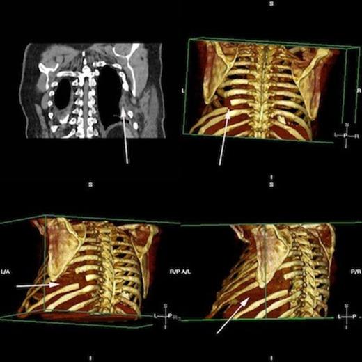
A CXR scan was performed which illustrated atelectasis at his left lung base. A subsequent CT demonstrated intercostal lung herniation alongside fractured sixth, seventh and eighth ribs, arising as a result of chest wall deformity (figure 1). Images were subsequently reconstructed by a clinical radiologist to fully demonstrate the skeletal pathology. (figure 2)
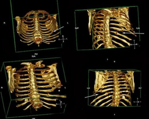
Rendered images of fractured ribs 6, 7 and 8 Arrowheads demonstrating fractures of the left 6th, 7th and 8th ribs. * = site of skeletal defect and subsequent site of pulmonary herniation
Being an exceptional case, a multi-disciplinary meeting was held with consulting members including the operating surgeon, a clinical radiologist, senior ward staff and a specialist respiratory physiotherapist. With respect to the severity of his chest pain, he was rendered unfit for work and with the resulting intrusion upon his quality of life, he was thus listed for surgical repair of the pathology. Unfortunately, surgery was delayed by six weeks due to the diagnosis of a lower limb deep vein thrombosis.
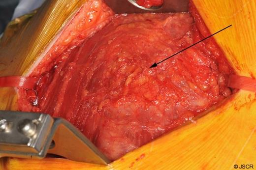
A limited left muscle sparing postero-lateral thoracotomy was used to gain access to the chest. Both the serratus anterior and latissimus dorsi muscles were mobilised and preserved (figure 3). The fractured portions of the seventh and eighth ribs were dissected and subsequently resected. The sixth rib, deemed to be healing appropriately and in-keeping with normal anatomy, was spared.
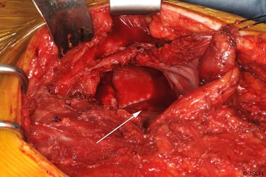
Next, the site of the lung herniation was identified (figure 4) and interrupted anchoring sutures were lain down (figure 5).
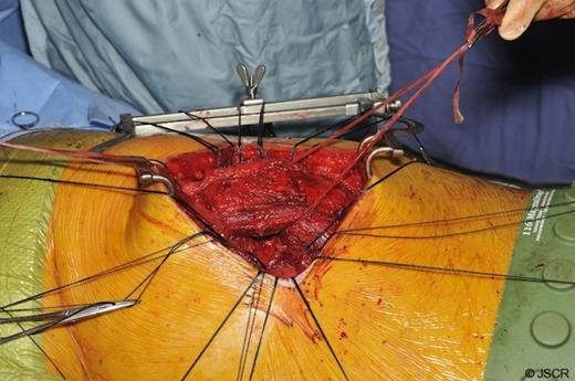
A 15 by 20cm, 1.5mm thick PermacolTM (Covidien, Hampshire, UK) biological implant was used to close the defect (figure 6 and 7). The soft tissues were then reinforced using interrupted sutures. The ribs that had been divided following the excision of the non union fragments were then approximated to restore a more normal intercostal space with interrupted peri-costal sutures. Haemostasis was achieved and a single 28F chest tube was placed within the pleural cavity.
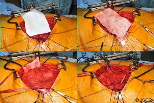
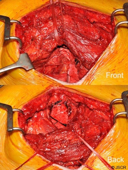
The PermacolTM bioprosthesis (Anterior and Posterior view) Latissimus Dorsi is reflected posteriorly and anteriorly respectively
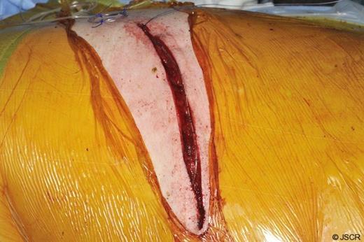
Finally a subcutaneous wound catheter was sited for post-operative control of the patient’s pain (figure 8). Clips were applied to the skin.
Postoperative progress was satisfactory and without complication, with the patient receiving daily input from the surgical, anaesthetic and physiotherapy teams. A repeat CT scan following the procedure confirmed restoration of the hernial defect and correction of the pulmonary hernia (figure 9).
Postoperative rendered images confirming restoration of herniated lung Top left, CT demonstrating PermacolTM patch in situ. Rendered images display areas of resected ribs and the revised bony form. Lung is restored and shows no evidence of herniation.
By the second postoperative week, the patient was mobile and pain free and subsequently discharged with outpatient follow-up. Subsequent clinic reviews revealed that although the patient was suffering with some neuropathic pain, his pre-operative symptoms had since resolved.
DISCUSSION
Lung herniation is a rare thoracic pathology. Repair is warranted as failure to treat can lead to subsequent atelectasis, chronic pain, deformity and restriction of pulmonary function (4). Rib fixation is infrequently described in the literature and there is no consensus on long term outcomes (2). There is some suggestion that early fixation can lead to an expeditious discharge, however there is minimal data to confirm this (2).
There are a variety of surgical interventions described to date and these include the use of wires, soft synthetic patches, Judet and sliding staples (3).
With the use of a soft prosthetic patch, intercostal space movement is facilitated with considerably less restriction upon mechanics than that of a rigid application such as the organic methylmetacrylate or other metal implants. Furthermore a bio-prosthetic implant confers a relatively reduced risk of infection whilst ensuring sustained durability.
We have described the first documented method for the use of a biological implant in the management of lung herniation.


