


Abstract
Epidermal cysts are very common lesions. Here we present the unusual case of an epidermal cyst occurring between the anal sphincters, presenting as a lump in the perineum. This was successfully excised with careful dissection of the intersphincteric plane. To our knowledge this is the only case of its kind reported in the literature.
INTRODUCTION
Epidermal cysts are common. They occur most frequently on the head and neck (1) but can arise anywhere on the body. Here we present an unusual case of an epidermal cyst arising in the intersphincteric plane of the anal canal. To our knowledge this is the only case of an epidermal cyst in this location reported in the literature.
CASE REPORT
A 41-year-old man presented with a 3 year history of a lump in the perineum. The lump had been steadily increasing in size over this time period, and had become uncomfortable. The patient was otherwise fit and well at presentation and there was no history of trauma to the perineum. His past medical history included a left orchidectomy for a seminoma 2 years previously. On examination, the lump was situated anterior to the anal canal. The lump was oval in shape, soft, smooth and did not appear to be infected. On digital rectal examination it could be felt extending superiorly. At the time of presentation it measured 60mm in maximal diameter.
Further characterisation using magnetic resonance imaging (MRI) revealed a very well defined mass returning high signal on T2 weighted images suggesting fluid or proteinaceous contents. The lesion appeared to originate from between the internal and external anal sphincters, with the superior part of the lesion therefore lying in the intersphincteric plane and the lower part bulging out into the perineum. (Figure 1)
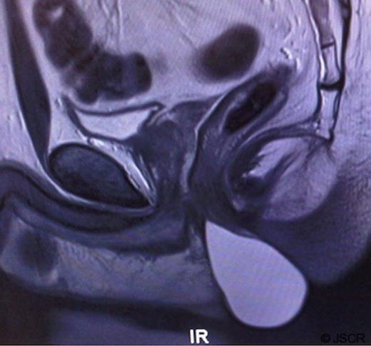
MRI of the pelvis showing a large cystic mass arising from the intersphincteric plane, lying anterior to the anal canal.
With the patient in the lithotomy position the lesion was excised in its entirety using careful dissection between the anal sphincters. Due to the size of the cavity left behind the skin was closed over a drain which was removed on the first post-operative day. The patient made a good recovery with no complications, including clinically intact anal sphincter function. To date there has been no recurrence. (Figure 2, Figure 3)
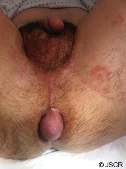
Pre-operative image of the perineum
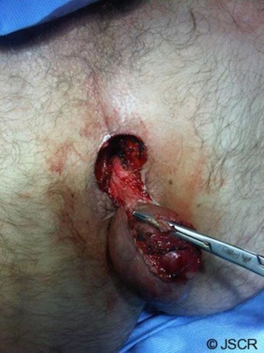
Intraoperative image of perineum
The macroscopic appearance was a cystic lesion with a 1mm thick wall containing brownish fluid. It measured 60mm x 30mm in maximum dimensions. Microscopically the cyst had a keratinizing squamous epithelium with no atypical features. The histological conclusion was an epidermal cyst.
DISCUSSION
Epidermal cysts are slow growing benign lesions which originate from the epidermal layer of the skin. They occur as a result of the proliferation of epidermal cells within the dermis and hence are often called epidermal inclusion cysts. Of note, there are a number of other forms of epidermoid type cysts also named according to the origin of the proliferating cells. In fact, the most common of all epidermoid cysts are follicular infundibular cysts which arise from cells originating in the upper portion of a hair follicle. Epidermal cysts do not usually cause symptoms, but they can cause discomfort either due to their size or if they become infected.
Skin trauma and inflammation have been implicated in the aetiology of epidermal cysts. Ultraviolet light exposure, tissue trauma (including surgery), smoking and human papilloma virus have all been linked with their development.(2-4) Epidermal cysts are also common in Gardener syndrome, an autosomal dominant condition which is a variant of familial adenomatous polyposis. There are occasional reports of malignant lesions arising from epidermal cysts although this is exceedingly rare.(5,6) In this case we could not identify any precipitating factors and, in particular, there was felt to be no link to the previous seminoma.
Differential diagnosis for a cystic lesion in the perineum includes a tail gut cyst (a developmental lesion lined with gastrointestinal epithelium which would be retro-rectal (7), perineal median raphe cysts (8) or urethral and seminal vesicle cysts which would be connected to the urological system. The clinical picture did not fit that of an abscess, and a lipoma was ruled-out due to the clearly cystic nature of the lesion on MRI.
There have been many reports of unusual sites for epidermal cysts which include intracranial, breast and para-rectal.(9,10) Epidermal cysts arising in the pelvis however are rare, with less than 10 case reports in the literature. Most of these cases are retro-rectal. This case is made even more unusual by the position of the cyst between the two anal sphincters. We have shown that in a case such as this, careful excision by an experienced surgeon can be safely and successfully undertaken with minimal morbidity.
