


Abstract
Appendicitis is a common surgical condition with various clinical presentations. The diagnosis could be obscured by underlying undiagnosed anatomical anomalies like intestinal malrotation. Intestinal malrotation is a rare foetal anomaly resulting from an incomplete, or failure of midgut rotation and fixation. 85% of cases have been estimated to present in the first two weeks of life. Presentation at adulthood is rare. In cases where peritonism is elicited elsewhere other than the right iliac fossa, clinicians should bear in mind the possibility of underlying intestinal malrotation, as this could be the first presentation of this rare congenital condition.
INTRODUCTION
Appendicitis is a common surgical condition. Its symptomatology could mimic other disease processes such as cholecystitis, diverticulitis, ruptured ovarian cysts and pelvic inflammatory disease. The diagnosis could be further obscured by underlying undiagnosed anatomical anomalies, for instance intestinal malrotation. Midgut malrotation is a foetal anomaly resulting from the failure of rotation around the axis of the superior mesenteric artery and subsequent retroperitoneal fixation. It is a rare condition with an approximate incidence of one in 500 live births (1). 85% of cases have been estimated to present in the first two weeks of life (2). Presentation at adulthood is rare. Hence, it is difficult to establish an accurate incidence as this condition can be asymptomatic and undetected until it is discovered in investigations or surgery for other abdominal pathologies in adulthood (3). Clinical suspicion of intestinal malrotation declines with age because of the rarity of the disorder. We report a case of perforated appendicitis in a middle-aged man with an incidental finding of midgut malrotation at the time of diagnostic laparoscopy.
CASE REPORT
A previously fit and well 56 year old gentleman presented to our emergency department with a two-day history of constant, dull, diffuse central abdominal pain. The pain worsened and became more localised to the epigastrium and left upper quadrant. The pain was associated with nausea and vomiting. He had no significant past medical history but he had a positive family history of colorectal carcinoma. He is a non-smoker and drinks 20 units of alcohol per week.
On examination, his observations were T36.7C, pulse 53 beats per minute and blood pressure of 120/75mmHg. On, palpation, tenderness with guarding, rebound and rigidity was elicited over the epigastrium and left upper quadrant. His biochemical profile showed raised inflammatory markers white cell count of 12 x109/L, neutrophils of 8.9 x10⁹/L, CRP of 62 mg/L and amylase of 40 U/L. Erect CXR was normal and showed no free air under diaphragm. Abdominal film was unremarkable.
Based on his worsening of symptoms and the clinical findings, emergency diagnostic laparoscopy was performed. The findings included a large amount of free pus in the peritoneal cavity, absence of appendix and caecum in the right lower quadrant and significant adhesions in left upper quadrant. After several attempts in dividing adhesions in the left upper quadrant, the decision was made to convert to open laparotomy. At laparotomy, the caecum and a perforated gangrenous appendix was found in the left upper quadrant as shown on the picture in Figures 1 and 2.
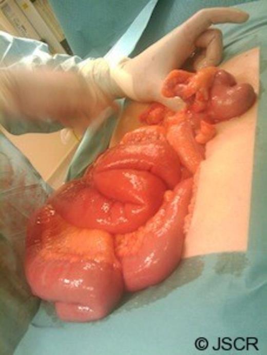
Operative image showing a perforated gangrenous appendix found in the LUQ
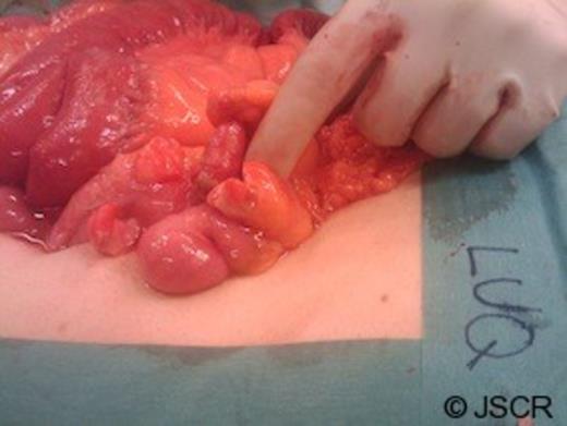
Operative image showing a perforated gangrenous appendix found in the LUQ
A malrotation at the jejunal level was found which attributed to the condition. Appendicectomy was performed with the caecum left at the left upper quadrant. Post-operatively, the patient had paralytic ileus and mild wound infection which resolved with conservative management and discharged on the 7th day.
DISCUSSION
Intestinal malrotation occurs in early foetal development. It is the failure of the normal 270° counterclockwise rotation of the midgut. The process occurs between the fourth and twelfth week of foetal life. At four weeks, the embryological gut exists as a single tube, suspended anterior to the superior mesenteric artery (SMA). The midgut enlarges rapidly beyond the capacity of the peritoneal cavity, herniating into the umbilical cord at six weeks. Within the cord, it rotates 90° counterclockwise around the axis of the SMA. This brings the third and fourth parts of the duodenum across to the left of the midline, behind the superior mesenteric artery. The mid-gut returns to the abdomen at the tenth and twelfth week. During this time, it continues to rotate a further 180° counterclockwise, bringing the ascending colon to the right side of the abdomen with the caecum lying immediately below the liver. The caecum descends into its normal position in the right iliac fossa, pulling the colon with it. Subsequent fusion and anchoring of the mesentery occurs. The third and fourth part of duodenum is fixed in the retroperitoneum, supported by the ligament of Treitz. The caecum is bound to the lateral abdominal wall by peritoneal bands. When any of the above sequent of events fails, the duodenojejunal flexure may freely hang from the foregut, at the right side of the abdomen. The caecum may be abnormally mobile or lying in the left upper quadrant as seen in our case report (4). This may result in dangerous conditions like intestinal obstruction or intestinal ischaemia of secondary to volvulus.
Diagnosis of intestinal malrotation is often an incidental finding in barium studies, angiographies, and computer tomography scans as found in various literature (5). Laparoscopy has a reputable value in the evaluation of atypical or undiagnosed abdominal pain. Presently, no reported cases of laparoscopically-diagnosed intestinal malrotation is found in the literature.
In conclusion, appendicitis is a common surgical condition with various clinical presentations. In cases where peritonism is elicited elsewhere other than the right iliac fossa, clinicians could bear in mind the possibility of underlying intestinal malrotation, as this could be the first presentation of this rare congenital condition.
