


 https://orcid.org/0009-0008-8484-9314

Abstract
Intestinal obstruction due to a giant fecaloma is a rare clinical condition, characterized by a markedly hardened fecal mass that is more severe than a simple fecal impaction. We report a case of a 24-year-old female with a history of repaired anorectal malformation (ARM) during infancy who presented with the complaint of abdominal rigidity, progressive abdominal distention, and chronic constipation. The contrast-enhanced computed tomography of the abdomen showed a grossly dilated rectosigmoid region. During laparotomy, a giant fecaloma (4 kg) was found obstructing the descending colon. The distal sigmoid stump was closed, and a diversion transverse colostomy was created to allow the passage of stool. The postoperative period was uneventful, and she was discharged with a well-functioning stoma. This case highlights that exploratory laparotomy with enterotomy and fecaloma extraction with the creation of a diversion transverse colostomy can be an effective and safe treatment approach, leading to excellent postoperative recovery and symptom relief.
Introduction
Chronic constipation leading to fecal impaction is a common condition, and its most severe form is a fecaloma, a hardened mass that can sometimes mimic the clinical and radiological appearance of carcinoma [1, 2]. While these masses typically localize in the rectosigmoid region, they rarely occur in the proximal colon or cecum [3]. Many studies have reported that fecalomas are prevalent in the elderly and debilitated population [4, 5]. However, an often overlooked high-risk group involves adult survivors of congenital anorectal malformations (ARM). Despite neonatal surgical correction, these patients frequently suffer from long-term motility problems and stricture formation leading to obstructive symptoms [6].
Case presentation
A 24-year-old female with subclinical hypothyroidism and dimorphic anaemia presented with a history of severe constipation and an inability to pass stool for the past 10 months, associated with progressive abdominal distension. She underwent anoplasty at 15 days of life for anorectal malformation, following which her family had been advised to perform daily anal dilatation using cervical dilators; however, they were inconsistent with it. Since then, she has experienced intermittent episodes of chronic constipation, which she managed with polyethylene glycol (PEGLEC) and occasional enemas. She was on 25 micrograms of thyroxine daily.
Earlier, she had been evaluated at a previous center, where an abdominal examination revealed a large, firm lump. Colonoscopy could not be performed due to massive fecal loading. A contrast-enhanced computed tomography of the abdomen performed there showed mild anal canal wall thickening (11.5 mm) with significant luminal narrowing, marked upstream dilatation of the rectum, sigmoid colon, and large bowel packed with fecal matter, and displacement of the urinary bladder and uterus by the distended rectum, findings suggestive of an anal stricture with obstruction. She was managed conservatively but showed no clinical improvement, so she was referred for definitive management.
On presentation, she was hemodynamically stable. Abdominal examination revealed a large, firm, well-defined mass predominantly on the left side of the abdomen with no guarding or peritoneal signs. After all the routine investigations were done, the patient was taken up for emergency surgery. During exploratory laparotomy, the descending colon was found filled with ~4–5 kg of hard fecaloma, which was manually evacuated (Figs 1 and 2). A portion of the descending colon was resected, and a diversion transverse colostomy was performed to allow the passage of stool. The distal sigmoid stump was closed. She was mobilized and started receiving chest physiotherapy on postoperative Day 1. She was kept nil by mouth until 48 hours and then was gradually allowed oral intake. An enema was administered through the stoma on postoperative Day 2, restoring its function. Osmotic laxative therapy with PEGLEC was given on POD 4 to increase the stoma output. The rest of the postoperative period was uneventful, with satisfactory stoma function and no postoperative complications. The patient was observed for the next 4 days and was discharged in stable condition on POD 8 with appropriate stoma care instructions.
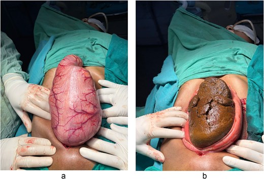
(a, b) Intraoperative view during exploratory laparotomy showing a grossly dilated and thinned descending colon containing a large, hardened mass.
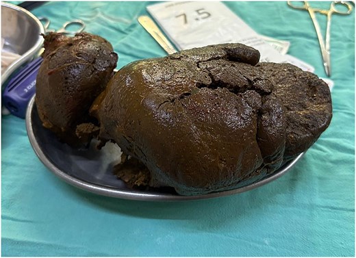
The extracted giant fecaloma, weighing ~4 kg, following manual evacuation and segmental resection of the descending colon.
On a 4-week follow-up, the patient was clinically stable with satisfactory stoma function and adequate output. She reported complete relief of abdominal distension and constipation, with no abdominal pain or other gastrointestinal complaints. No stoma-related complications were observed. A further management plan, including the possibility of stoma reversal, will be guided by serial clinical assessment of the distal bowel patency and anorectal outlet.
Discussions
Etiology and pathophysiology
Fecaloma is a cycle of chronic constipation in which the colonic mucosa continues to absorb water from the fecal matter, leading to progressive hardening and lamination of the stool [1]. Apart from motility issues in elderly, debilitated, and bedridden patients, the underlying causes of fecaloma are diverse. A retrospective study by De Souza DH et al. observed that destruction of the intramural nerve plexus due to Chagas disease, leading to megacolon, subsequently results in fecaloma formation [7]. The etiology in younger patients is often different (Table 1). For instance, the use of anticholinergics or antipsychotic medications for schizophrenia has been shown to severely impair colonic motility and, if neglected, can lead to massive fecalomas, some reaching up to 30 cm in diameter [9]. Our case presents a distinct and significant etiology, a congenital ARM. This highlights that fecaloma must remain a primary differential diagnosis in any patient with a history of chronic constipation, regardless of age or previous surgical interventions [10]. Unlike most reported giant fecalomas, which occur in elderly, debilitated, or psychiatrically medicated patients [1, 5, 8, 9], our case involves a young adult survivor of repaired ARM, representing the under-recognized group in whom fecaloma formation is driven by anal stenosis and long-standing dysmotility.
Etiological classification and pathophysiological mechanisms of fecaloma
| Category | Causes/risk factors | Mechanism |
|---|---|---|
| Congenital or anatomical |
| Agangliogenesis leading to aperistalsis [7] Functional or mechanical obstruction [8] |
| Pharmacological |
| Decreased intestinal motility and increased water absorption [9] |
| Neurological and psychiatric |
| Destruction of the intramural nerve plexus [11] Neglect of the urge to defecate |
| Metabolic and endocrine | Hypothyroidism Diabetes Chronic renal failure Hypercalcemia | Metabolic slowdown affecting smooth muscle contractility is common, but leading to fecalomas is extremely rare [7] |
| Lifestyle and others | Low-fiber diet Chronic dehydration Sedentary lifestyle (elderly, debilitated, or bedridden) | Formation of small, hard stools that coalesce into a laminated mass [7] |
The risk of complications
Management decisions depend crucially on the risk of developing life-threatening complications in patients with fecaloma. As the mass grows, it exerts direct pressure on the bowel wall and surrounding structures, leading to stercoral ulceration, pressure necrosis, and gastrointestinal bleeding. In the most severe cases, it can progress to intestinal perforation and peritonitis, which potentially have high mortality rates [10].
The sheer size of these masses can often cause mass effects on adjacent organs. There are several studies that reported fecaloma causing urinary outlet obstruction, leading to hydronephrosis and acute renal failure [12]. While our patient presented with bladder displacement, she did not manifest obstructive symptoms, unlike other reports, emphasizing the importance of intervention before such multisystem complications arise.
Management
The size of the mass, its location, and the presence of complications determine the management strategy for fecaloma. For smaller or distal fecalomas, conservative measures such as enemas or manual disimpaction are the first-line treatment [10, 13]. Some reports have shown the use of endoscopic fragmentation with specialized snares or even chemical dissolution when routine methods fail [14, 15]. Surgery is indicated when the conservative and endoscopic management fails, when the mass is in the proximal colon, or when complications like perforation are suspected. Our patient, with no complications, presented with a 4 kg mass in the descending colon. This condition was also far beyond the reach of endoscopic therapy. The choice between a simple enterotomy and a formal resection depends on clinical judgment. In our case, because the descending colon was chronically thinned and dilated, a resection with transverse colostomy diversion was performed.
Long-term bowel surveillance is important in ARM survivors. Despite technically successful neonatal repair, these patients have an increased risk for anal stricture, chronic constipation, and dysmotility, which can manifest as obstruction anytime in life [16]. Structured bowel management, consistent adherence to anal dilatations, and periodic clinical and endoscopic review are essential to prevent stool stasis and fecaloma formation. In our patient, inconsistent dilatations during childhood likely contributed to the development of a stricture and an eventual obstructive presentation in adulthood, reinforcing the need for sustained follow-up.
In conclusion, a giant fecaloma is a rare but life-threatening complication, particularly in adult survivors of anorectal malformations. While conservative management is the first choice, supergiant fecalomas often necessitate surgical intervention. This case demonstrates that exploratory laparotomy and diversion colostomy are safe and effective procedures for fecalomas in preventing complications and allowing a stable postoperative recovery. Long-term monitoring of bowel habits in ARM patients is essential to prevent extreme sequelae.
Author contributions
Dinesh Choudhary (Idea, Conceptualization, Supervision, Writing draft, Approved final draft), Gunvi Ohri (Idea, Conceptualization, Supervision, Writing draft, Approved final draft), Shivani Ravipati (Resources, Formal analysis, Writing draft and Revision of draft, Approved final draft), Pranav Vempati (Resources, Formal analysis, Writing draft and Revision of draft, Approved final draft), Bhaskar Kambhampati (Resources, Formal analysis, Writing draft, Revision of draft, Approved final draft), and Tejinder Singh (Conceptualization, Project administration, Formal analysis resources, Writing, Revision, Approved final draft)
Conflicts of interest
The authors declare no conflict of interest.
Funding
No funding received.
Data availability
Not applicable.
Ethics statement
In our university, ethics approval was not required for case reports and case series.
Informed consent
Consent was obtained from the patient for the research and the publication.


