


 https://orcid.org/0000-0001-7754-4629

Abstract
The finding of an adrenal mass in a patient with a history of extra-adrenal malignancy represents a diagnostic dilemma, in which metastasis is usually the primary suspicion. Pure adrenal lipomas are extremely rare benign tumors (<5% of adrenal neoplasms) that can coexist with other malignancies. We present the case of a 66-year-old female with a history of infiltrating ductal carcinoma of the breast treated in 2023. During her surveillance protocol, a computed tomography scan revealed a right renal mass and an ipsilateral adrenal lesion with fat density. Given the clinical suspicion of a second primary renal tumor and possible metastatic activity, a right radical nephrectomy was performed. Histopathological analysis confirmed a high-grade clear cell renal cell carcinoma (Grade 4, sarcomatoid features) pT1a, and a synchronous pure adrenal lipoma, definitively ruling out metastasis and myelolipoma. This case underscores the importance of an accurate radiological-pathological correlation to avoid diagnostic bias in complex oncological patients.
Introduction
The finding of adrenal incidentalomas has increased exponentially due to the widespread use of computed tomography (CT) and magnetic resonance imaging, being reported in up to 5% of the general population [1]. However, the detection of an adrenal mass in a patient with a history of extra-adrenal malignancy poses a much more complex clinical scenario.
Recent studies indicate that, in oncological patients, between 30% and 70% of incidental adrenal masses correspond to metastases, with the most common primary sites being the lung, kidney, and breast [2, 3]. Among benign lesions, tumors with macroscopic fat content are usually diagnosed as myelolipomas. Conversely, the pure adrenal lipoma (a benign mesenchymal neoplasm composed exclusively of mature adipocytes without hematopoietic elements) is an extremely rare entity, with a reported incidence of less than 0.7% of all adrenal tumors [4, 5].
The synchronous coexistence of a clear cell renal cell carcinoma (ccRCC), a history of breast cancer, and an ipsilateral adrenal tumor constitutes an unusual clinical triad. The objective of this report is to present the management of this case and discuss the critical radiological and pathological characteristics essential for the differential diagnosis.
Case report
We present the case of a 66-year-old female with a history of systemic arterial hypertension and uncomplicated diverticular disease. In 2023, she was diagnosed with breast carcinoma of no special type, Nottingham histologic grade 2. Immunohistochemical analysis classified the tumor as luminal B, with cytological confirmation of axillary lymph node metastasis (positive fine-needle aspiration biopsy for carcinoma). She was treated with a mastectomy and adjuvant therapy, remaining under surveillance without evidence of local recurrence.
During oncological follow-up in 2024, a CT scan revealed a space-occupying lesion in the right kidney (Fig. 1a and c) suggestive of a second primary tumor, in addition to indeterminate pulmonary and abdominal lymphatic findings that raised the possibility of metastatic tumor activity. A hypodense nodular lesion was concomitantly identified in the right adrenal gland with a density of −99 Hounsfield units (HU) (Fig. 1b and d), suggesting an adipose composition.
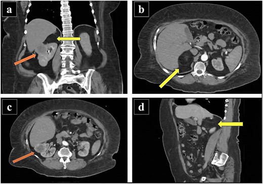
Contrast-enhanced abdominal CT. (a) Coronal image showing a solid lesion in the upper pole of the right kidney, corresponding to clear cell renal cell carcinoma, and an ipsilateral right adrenal lesion. (b) Axial image demonstrating a fat-containing lesion in the right adrenal gland, compatible with a lipoma. (c) Axial image showing the solid lesion in the upper pole of the right kidney, corresponding to clear cell renal cell carcinoma. (d) Sagittal image demonstrating the right adrenal lesion, compatible with a lipoma. Arrows indicate the respective renal and adrenal lesions.
In 2025, the patient was admitted for definitive surgical management. Preoperative laboratory tests were within normal parameters; therefore, an open right radical nephrectomy was performed. Surgical findings included a right kidney measuring 12.0 × 8.0 × 7.0 cm with a neoformation in the posterior pole, as well as an ipsilateral adrenal gland displaying a well-demarcated, yellowish nodular appearance. The surgical technique involved the dissection of the renal hilum, identifying two renal arteries and one main renal vein. En bloc excision was successfully completed.
Macroscopic examination of the right adrenalectomy specimen (measuring 6.0 × 5.0 × 4.0 cm and weighing 82 g) revealed a smooth, grayish-brown outer surface. Serial sectioning exposed a solid, yellowish, adipose-like tumor with reddish hemorrhagic areas (Fig. 2a and b).
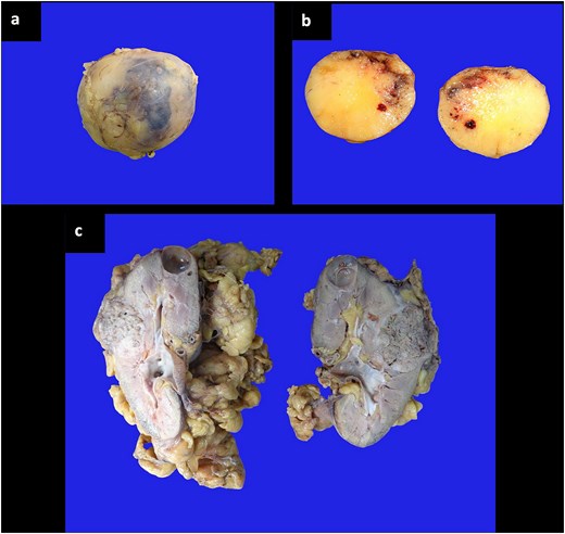
Macroscopic examination showing (a, b) right adrenalectomy specimen with a solid, yellowish tumor of adipose appearance, (c) radical nephrectomy specimen with the presence of a tumor in the middle third and a cyst in the upper pole.
The radical nephrectomy specimen, received in a separate container, measured 16.0 × 9.0 × 5.5 cm and was surrounded by abundant soft, yellowish adipose tissue. Dissection revealed a kidney measuring 11.5 × 6.5 × 4.5 cm with a grayish-brown, poorly defined tumor in the middle third (4.0 × 3.2 cm) featuring myxoid areas. The tumor pushed against, but did not perforate, the renal capsule. The residual corticomedullary ratio was preserved. A simple cyst (1.5 × 1.8 cm) with smooth internal walls was identified in the upper pole (Fig. 2c).
Histopathological analysis reported a malignant neoplasm of epithelial lineage characterized by marked heterogeneity. The conventional clear cell component consisted of nests of cells with optically empty cytoplasm and well-defined cell membranes, immersed in a delicate capillary network (Fig. 3a). This transitioned into a sarcomatoid component characterized by spindle-shaped cells arranged in fascicles (Fig. 3b). Concomitantly, rhabdoid differentiation was identified, manifested by large, discohesive cells with markedly eosinophilic cytoplasm, eccentrically placed nuclei, open chromatin, and prominent nucleoli (Fig. 3c). No lymphovascular invasion was observed. The adrenal gland contained an encapsulated nodule of 6.0 cm in its longest axis, composed exclusively of mature adipose tissue and negative for hematopoietic elements, confirming the diagnosis of a pure adrenal lipoma (Fig. 3d).
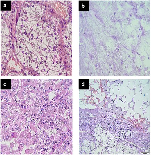
Histological examination showing (a) classic clear cell renal cell carcinoma, (b) sarcomatoid differentiation, (c) rhabdoid differentiation, and (d) pure adrenal lipoma.
The patient had a favorable postoperative evolution and was discharged on the third postoperative day. During her long-term follow-up, she has demonstrated an excellent clinical evolution, maintaining an Eastern Cooperative Oncology Group performance status of 0. All follow-up laboratory and imaging studies have remained within normal limits, with no evidence of tumor recurrence or distant disease. Consequently, she was formally discharged by the Urology and Surgical Oncology departments, and continues exclusively under a strict surveillance protocol by the Medical Oncology department.
Discussion
The evaluation of an adrenal mass in the context of a known malignancy requires ruling out metastasis through imaging before considering biopsy [1, 6]. In our case, the diagnostic challenge lay in the patient’s history of metastatic breast cancer. Although the negative attenuation values on CT, strongly suggested fat (benignity), the surgical decision was driven by the presence of the synchronous renal tumor and the impossibility of ruling out malignancy with absolute certainty without histological confirmation.
The definitive diagnosis of pure adrenal lipoma is strictly histological. It is differentiated from a myelolipoma by the complete absence of hematopoietic tissue (myeloid/erythroid lineages) and positive S-100 staining in the adipocytes [4, 7].
From an oncological perspective, this case is highly illustrative. Clinically, an advanced stage (cT3a or metastatic disease) was suspected. The final pathology reclassified the patient to Stage I (pT1a), drastically improving her prognosis. However, the finding of sarcomatoid and rhabdoid differentiation (WHO/ISUP Grade 4) is an independent poor prognostic factor regardless of tumor size, mandating close surveillance despite the early pT1a stage.
The en bloc radical nephrectomy not only effectively treated the aggressive renal cancer but also resolved the diagnostic uncertainty surrounding the adrenal mass, unequivocally confirming its benign nature.
Currently, there are no published studies reporting the synchronous presence of these three specific entities in the same patient linked to known predisposition syndromes. However, the literature indicates that mutations in the PTEN gene increase the risk of both breast cancer and clear cell renal cell carcinoma, and the loss of PTEN function has been identified as a direct cause of lipoma formation in the adrenal cortex [8].
Conclusion
The coexistence of a pure adrenal lipoma and a renal carcinoma with sarcomatoid differentiation in the context of a history of breast cancer constitutes an exceptional clinical scenario. This case demonstrates that negative radiological density is a reliable predictor of adrenal benignity, even in high-risk oncological patients. However, en bloc radical nephrectomy was validated not only as a definitive diagnostic tool but as a crucial oncological treatment for a renal carcinoma with aggressive biological behavior (Grade 4) that, despite its early stage (pT1a), carries a high risk of recurrence. The precise histopathological distinction reclassified the patient’s prognosis, transforming a presumably metastatic scenario into a locally controlled disease that warrants strict surveillance.
Conflicts of interest
The authors declare no conflicts of interest.
Funding
No funding was received for this study.
References
Bokhari MR, Zulfiqar H, Leslie SW et al. Adrenal myelolipoma. In:


