


 https://orcid.org/0000-0002-6827-0088

Abstract
Subhepatic appendicitis is a rare anatomical variant that mimics hepatobiliary pathology, leading to diagnostic delay and technical difficulty at surgery. We describe a 69-year-old man who underwent emergency laparoscopic appendicectomy for sepsis, complicated by dense subhepatic adhesions resulting in incomplete resection and a retained appendiceal tip. Histopathological examination unexpectedly demonstrated moderately differentiated appendiceal adenocarcinoma with isolated loss of postmeiotic segregation increased 2 expression. Three weeks later, the patient re-presented with recurrent sepsis due to pyogenic liver abscesses and a hepatic lesion suspicious for metastatic disease. Multidisciplinary assessment led to radical right hemicolectomy, cholecystectomy, and limited liver resection. Final histopathology confirmed locally advanced disease with direct T4 invasion into the liver and clear (R0) margins. This case highlights the need to consider subhepatic appendicitis in unexplained right upper quadrant sepsis and emphasizes the importance of early multidisciplinary management when retained appendiceal tissue is suspected, as radical resection may be curative.
Introduction
Subhepatic appendicitis is a rare anatomical variant, accounting for approximately 0.01%–0.08% of appendicectomies, most commonly resulting from abnormal embryological rotation or maldescent of the caecum [1]. Its atypical location often leads to misdiagnosis as hepatobiliary or renal pathology, resulting in delayed intervention and increased rates of perforation, abscess formation, and sepsis. Surgical management can be technically demanding due to distorted anatomy, dense adhesions, and proximity to the gallbladder, duodenum, and liver.
While laparoscopic appendicectomy offers improved visualization in atypical presentations, complete resection may not always be safely achievable in complex cases. Retained appendiceal tissue following appendicectomy is uncommon but clinically significant. Beyond recurrent infection, residual tissue may harbour occult malignancy, with important oncological implications [2]. We present a rare case of subhepatic appendicitis complicated by a retained appendiceal tip containing adenocarcinoma, leading to recurrent sepsis and a diagnostic dilemma involving suspected hepatic metastasis.
Case report
A 69-year-old male presented with a 4-week history of intermittent right upper and right lower quadrant abdominal pain associated with unintentional weight loss. Symptoms acutely worsened over 24 h, with fever (39.3°C) and signs of right upper quadrant peritonitis. Laboratory investigations demonstrated marked systemic inflammation (C-reactive protein 143 mg/l) with a normal white cell count. Contrast-enhanced computed tomography (CT) was equivocal, raising concern for either perforated subhepatic appendicitis (Fig. 1a and b) or perforated cholecystitis (Fig. 2). Abdominal ultrasound demonstrated a normal gallbladder but identified a 13-mm inflamed appendix and a 45-mm porta hepatis abscess.
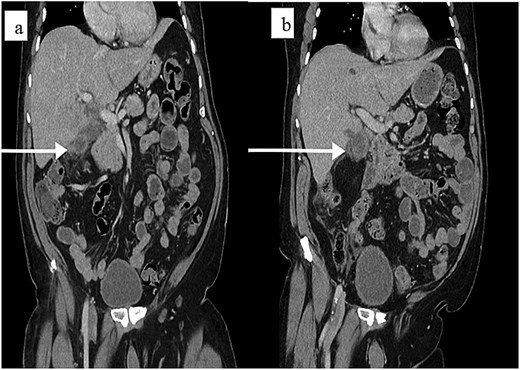
(a) Coronal contrast-enhanced CT of the abdomen and pelvis. The image demonstrates a rare subhepatic position of the appendix (arrow), appearing dilated, and inflamed tucked underneath the liver edge. (b) Surrounding inflammatory changes and localized fluid collection in the right upper quadrant, mimicking acute cholecystitis (coronal view).
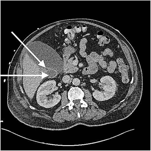
Axial contrast-enhanced CT demonstrating a distended, thin-walled gallbladder (horizontal arrow) in close proximity to a subhepatic inflammatory mass (diagonal arrow). The inflammatory changes from the subhepatic appendicitis mimic the presentation of acute cholecystitis, illustrating the diagnostic challenge of this anatomical variant.
Due to diagnostic uncertainty, initial conservative management was attempted; however, clinical deterioration necessitated operative intervention. Emergency laparoscopic appendicectomy revealed a subhepatic appendix densely adherent to the gallbladder and duodenum within severe inflammatory adhesions. Complete resection was not safely achievable, and a portion of the appendiceal tip was left in situ. The postoperative course was initially unremarkable. Histopathological examination of the resected specimen unexpectedly demonstrated moderately differentiated appendiceal adenocarcinoma with isolated loss of postmeiotic segregation increased 2 (PMS2) expression on immunohistochemistry.
Three weeks following discharge, the patient re-presented with sepsis (C-reactive protein 406 mg/l; white cell count 16 × 109/l). Repeat CT imaging demonstrated multiple pyogenic liver abscesses (Fig. 3a), which were managed with percutaneous drainage (Fig. 3b), and a solid lesion in hepatic segments 5/6 suspicious for metastatic disease (Fig. 4). Following multidisciplinary team discussion involving colorectal surgery, hepatobiliary surgery, oncology, radiology, and pathology, the patient underwent radical right hemicolectomy, cholecystectomy, and limited liver resection after a Positron Emission Tomography (PETscan) (Fig. 4). Final histopathology confirmed moderately differentiated adenocarcinoma with direct invasion into the hepatic parenchyma (T4 disease) and clear resection margins (R0). No distant metastatic disease was identified. Surveillance colonoscopy and interval CT imaging at four months and one year demonstrated no recurrence or peritoneal disease.
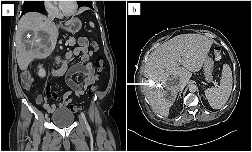
(a) Coronal contrast-enhanced CT of the abdomen performed three weeks postoperatively. The image demonstrates multiple large, multi-loculated pyogenic abscesses (asterisks) within the right lobe of the liver. These occurred secondary to recurrent sepsis from the retained appendiceal tip. (b) Resolution of abscess after insertion of percutaneous drain (axial view).
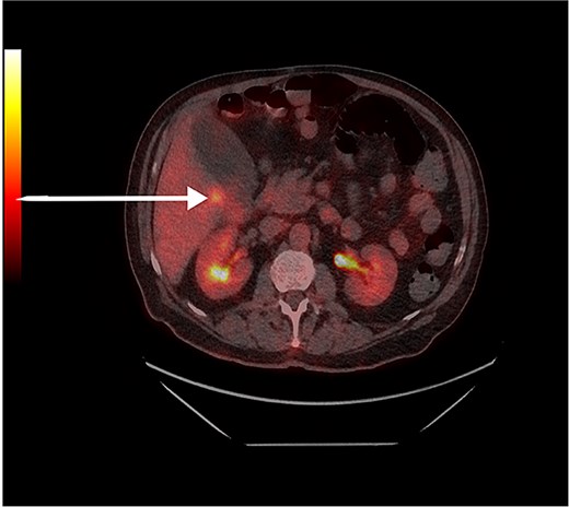
18F-fluorodeoxyglucose (FDG) PET/CT fusion image showing a focal area of intense hypermetabolism (arrow) within hepatic segment 5/6. This metabolic activity is characteristic of the patient's appendiceal adenocarcinoma with direct hepatic invasion (T4 disease). Normal physiological tracer uptake is noted in both kidneys (axial view).
Discussion
Subhepatic appendicitis represents a diagnostic challenge due to its atypical presentation, frequently mimicking biliary pathology. Contrast-enhanced CT remains the gold standard for diagnosis, with reported sensitivity of 80%–100%, significantly outperforming ultrasound in atypical anatomical locations [2]. Persistent right upper quadrant pain refractory to standard biliary management should prompt consideration of appendiceal malposition.
Incomplete appendicectomy in complex anatomy is rare but carries significant clinical consequences. Retained appendiceal tissue can act as a nidus for ongoing infection, as demonstrated by this patient’s recurrent sepsis and pyogenic liver abscesses. Importantly, residual tissue may also harbour occult malignancy, emphasizing the need for careful intraoperative assessment and postoperative vigilance [2]. Appendiceal adenocarcinoma is identified in fewer than 0.2% of appendicectomy specimens [3]; however, over 10% of affected patients have germline variants associated with hereditary gastrointestinal cancer syndromes [4]. The isolated loss of PMS2 expression in this case indicates mismatch repair deficiency and warrants evaluation for Lynch syndrome, with implications for both patient management and familial screening [4].
The presence of a solid hepatic lesion created a significant diagnostic dilemma, requiring differentiation between metastatic disease and direct invasion. In subhepatic appendicitis, close anatomical proximity facilitates direct extension into the liver. According to the American Joint Committee on Cancer (AJCC) 9th edition staging system, such invasion constitutes locally advanced (T4) disease rather than distant metastasis [5]. Achieving an R0 resection in T4 appendiceal adenocarcinoma is an independent predictor of long-term survival and offers a potential for cure not afforded in metastatic disease [6, 7]. Systematic reviews support radical right hemicolectomy over simple appendicectomy to achieve adequate lymphadenectomy and oncological clearance in appendiceal adenocarcinoma [8].
Conclusion
Subhepatic appendicitis is a rare but important diagnostic masquerade. Incomplete appendicectomy in atypical anatomy carries substantial risks, including recurrent sepsis and missed occult malignancy. This case highlights the importance of meticulous surgical assessment, routine histopathological review, and early multidisciplinary involvement when retained appendiceal tissue is suspected. Radical en-bloc resection with clear margins can achieve excellent oncological outcomes even in the presence of hepatic involvement abnormally positioned.
Learning points
Subhepatic appendicitis is a rare anatomical variant that frequently mimics hepatobiliary disease, leading to significant diagnostic and surgical challenges.
Incomplete resection of a subhepatic appendix may lead to recurrent sepsis and the potential for missed occult malignancy.
Direct hepatic invasion by appendiceal adenocarcinoma (T4 disease) should be differentiated from metastatic (M1) disease, as radical R0 resection can still offer curative potential.
Isolated loss of PMS2 expression in appendiceal specimens warrants clinical evaluation for Lynch Syndrome.
Conflicts of interest
The authors do not have financial or non-financial interests to disclose.
Funding
No funding was received for conducting this study.
Data availability
The data that support the findings of this study are available on request from the corresponding author. The data are not publicly available due to privacy or ethical restrictions.
Consent
The patient provided informed consent for the publication of this case report.


