


 https://orcid.org/0009-0007-9498-3894

Abstract
Extralobar pulmonary sequestration (ELS) with torsion is rare in adolescents and mimics posterior mediastinal tumours, posing diagnostic challenges. An 11-year-old boy presented with acute chest pain. Contrast-enhanced computed tomography revealed a non-enhancing homogeneous mass with pleural effusion, while magnetic resonance imaging showed isointense T1 and hypointense T2 signals. Despite initial uncertainty regarding neoplasm, the acute presentation contrasted with the indolent course of posterior mediastinal tumours. History of abnormal prenatal ultrasonography facilitated diagnosis of torsed ELS. Video-assisted thoracoscopic surgery confirmed infarcted ELS with torsion, and the patient recovered uneventfully. This case highlights the importance of including ELS torsion in the differential diagnosis of posterior mediastinal masses and underscores the diagnostic value of prenatal imaging history.
Introduction
Pulmonary sequestration (PS) is a rare congenital lung abnormality divided into intralobar and extralobar (ELS) types based on pleural coverage [1]. While ELS is commonly detected antenatally, infants may present with respiratory distress or high-output congestive heart failure [2]. ELS with torsion is extremely rare, with only 25 cases reported in the English literature according to a 2025 literature review, and the inability to visualize feeding arteries on imaging increases diagnostic difficulty [3]. Herein, we report a case of ELS with torsion in an 11-year-old patient. This study was conducted in accordance with the Declaration of Helsinki and the Surgical CAse REport (SCARE) guidelines SCARE guidelines is a standardised method for reporting surgical cases in the medical literature) [4], and written informed consent was obtained from the patient’s legal guardians.
Case report
The clinical timeline is summarized in Table 1. An 11-year-old boy presented with a 2-day history of severe right-sided chest pain. The pain was persistent, non-radiating, worsened by deep inspiration, and partially relieved by breath-holding. Three days after symptom onset, he began to experience abdominal pain. Past medical history, physical examination, and vital signs were unremarkable. Outside-hospital computed tomography (CT) revealed a mass-like pleural lesion behind the posterior basal segment of the right lower lobe (46 × 19 mm; 45 HU), raising suspicion for a posterior mediastinal tumour. The patient was referred for further evaluation, and analgesics were administered during hospitalization.
The clinical timeline.
| Time Point | Clinical events |
|---|---|
| 2 Days Pre-admission | Child developed severe right-sided chest pain. |
| 1 Day Pre-admission | External CT scan showed pleural mass. |
| Admission Day | Complete history and laboratory tests, no significant abnormalities. |
| Post-Admission Day 1 | CT and MRI completed, tumour suspected based on imaging but inconsistent with symptoms. |
| Post-Admission Day 2 | Department discussion considered pulmonary sequestration with torsion, obtained prenatal ultrasound history showing intrathoracic abnormal echo, diagnosis confirmed. |
| Post-Admission Day 3 | Surgery scheduled, preoperative preparation completed. |
| Post-Admission Day 4 | Thoracoscopic surgery under general anesthesia, confirmed pulmonary sequestration with torsion, drainage tube placed. |
| Post-Admission Day 6 | Chest X-ray repeated, drainage tube removed,discharged on the following day. |
Laboratory evaluation provided no significant diagnostic clues. Contrast-enhanced CT (Fig. 1A) demonstrated a non-enhancing pleural-based oval mass in the posterior basal segment of the right lower lobe (30.1 × 47.0 × 2.8 mm) with crescent-shaped pleural fluid. The magnetic resonance imaging (MRI) (Fig. 2A and B) showed right pleural effusion (maximum depth 16.9 mm) and a fusiform paraspinal lesion, isointense on T1 and hypointense on T2, measuring 49.9 × 16.9 × 46.0 mm. Echocardiography was normal.
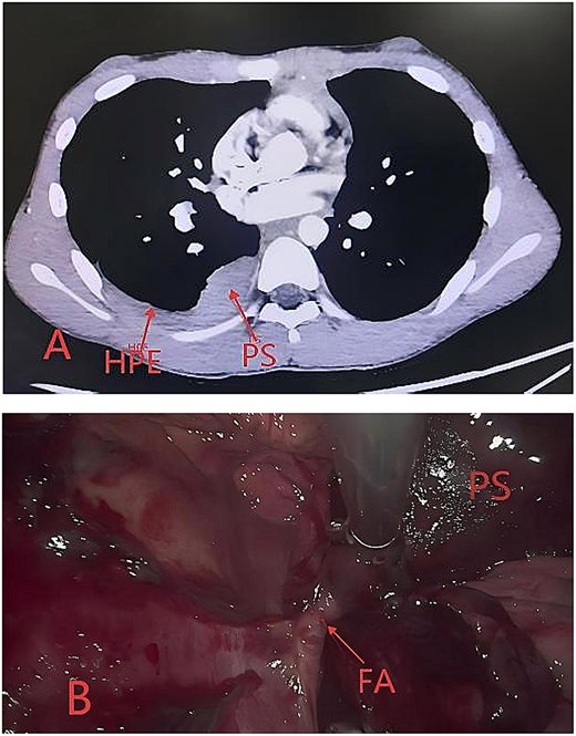
Contrast-enhanced CT findings of ELS with torsion and intraoperative images from video-assisted thoracoscopic surgery. Abbreviation: HPE (hemorrhagic pleural effusion), PS (pulmonary sequestration), FA (feeding artery). (A) ELS with torsion showed no enhancement on contrast-enhanced CT. (B) A feeding artery originating from the intercostal artery demonstrated a 360° rotation.
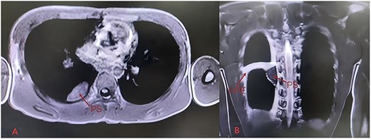
MRI findings of ELS with torsion abbreviation: HPE (hemorrhagic pleural effusion), PS (pulmonary sequestration). (A) T1-weighted imaging (isointense T1 signal); (B) T2-weighted imaging (hypointense T2 signal).
Based on a posterior mediastinal mass with pleural effusion, a posterior mediastinal tumour was initially suspected. However, the acute severe pain was atypical for pediatric posterior mediastinal tumours, which are usually asymptomatic or present with compressive symptoms due to mass effect rather than acute severe pain. CT and MRI demonstrated a well-circumscribed lesion without invasive features. Given the benign-appearing extrapulmonary lesion and our institutional experience as a regional pediatric center, ELS was included in the differential diagnosis. Nonetheless, contrast-enhanced CT did not identify a systemic feeding artery, and the lesion showed no enhancement. Moreover, ELS is typically asymptomatic and rarely associated with pleural effusion. To further clarify the diagnosis, the prenatal history was reviewed. Review of prenatal history revealed a hyperechoic mass in the right lower lobe at 24 weeks’ gestation, which appeared ‘disappeared’ later in pregnancy. A diagnosis of torsed ELS was therefore favoured, which can appropriately explain the clinical and imaging findings.
Thoracoscopic surgery was performed. Intraoperatively, a dark purplish mass was identified on the right posterior chest wall, measuring ~30 × 30 × 20 mm, with dense pleural adhesions. An aberrant feeding artery from an intercostal artery was twisted 360°clockwise (Fig. 1B). Histopathology confirmed ELS. The patient was discharged on postoperative Day 3 and remained asymptomatic at follow-up.
Discussion
ELS consists of a mass of lung parenchyma enclosed within its own pleural covering and is anatomically completely separated from the adjacent normal lung tissue. Although ELS has an independent pleural investment, it lacks the ligamentous attachments characteristic of normal lung tissue. This anatomical feature allows ELS to rotate around its systemic vascular pedicle, potentially resulting in torsion [5].
ELS with torsion usually presents in children and adolescents [3], although rare cases have been reported in infant (7-month) [6] and elderly patient (83-year) [3]. Most patients present with acute abdominal pain and vomiting, which may be attributed to diaphragmatic irritation caused by the typical location of ELS between the diaphragm and the lower lobe [3]. However, in the present case, the lesion was located away from the diaphragm, resulting in chest pain rather than abdominal pain. The abdominal pain that developed later in the clinical course was likely attributable to diaphragmatic irritation caused by the accompanying pleural effusion. The exact mechanism underlying torsion remains unclear. While vigorous physical activity has been suggested as a predisposing factor [5], the present patient had no history of strenuous activity prior to symptom onset.
Contrast-enhanced CT is the most commonly used technique for diagnosing ELS. But torsion may obscure the feeding vessel due to the lack of blood flow [7], increasing the difficulty of preoperative diagnosis. The contrast-enhanced CT findings of torsed ELS vary; some cases demonstrate irregular central enhancement with decreased peripheral enhancement [8], whereas others show only peripheral enhancement [9] or no enhancement [10]. In the present case, the absence of enhancement likely reflected complete infarction caused by 360°torsion of the feeding artery.
MRI findings also vary, with some cases demonstrating slightly high T1 and low T2 signal intensity [5] and others showing intermediate T2 signal intensity [11]. In the present case, MRI revealed isointense T1 and low T2 signal intensity without enhancement, findings suggestive of complete occlusion of the feeding vessel with complete infarction.
Radiologically, ELS with torsion presents as a posterior mediastinal mass. Due to its rarity and the lack of visible feeding arteries on imaging, differentiating ELS with torsion from posterior mediastinal tumour is challenging, which may lead to delayed treatment. In the present case, surgical treatment was performed on the sixth day after symptom onset due to an unclear preoperative diagnosis. The main distinguishing features between ELS with torsion and posterior mediastinal tumour that we learned from the case are as follows: posterior mediastinal tumours are usually asymptomatic or cause manifestations solely due to mass effect or compression, and typically demonstrate irregular margins on imaging. However, ELS with torsion is often accompanied by severe and obvious symptoms, and typically demonstrates well-defined margins with clear borders on imaging; Inquiry regarding prenatal ultrasonographic results is of great diagnostic value, as ELS frequently shows abnormal intrathoracic echogenicity on prenatal ultrasound.
Conclusion
ELS with torsion predominantly affects children and adolescents. Due to its rarity and the lack of visible feeding arteries on imaging, preoperative diagnosis remains challenging. However, characteristic clinical and imaging features may aid in differentiating this entity from neoplastic disease. Furthermore, the presence of abnormal intrathoracic echogenicity on prenatal ultrasound is of great value in confirming the diagnosis of ELS.
Conflicts of interest
None declared.
Funding
None declared.


