


 https://orcid.org/0000-0001-8843-9525

Abstract
Colonoscopy is generally considered a safe procedure, with extra-colonic complications being exceedingly rare. Gallbladder haemorrhage following colonoscopy has been reported only in isolated cases and remains a poorly recognized entity, particularly in patients with cirrhosis. We present a 52-year-old male with decompensated alcoholic cirrhosis who developed abdominal pain, anemia, and hypotension following an unremarkable colonoscopy. Initial imaging was inconclusive, and the patient was discharged after stabilization. He later re-presented with recurrent bloody ascites requiring multiple transfusions. Angiography identified a cystic artery pseudoaneurysm, which was embolized. However, the patient continued to require transfusions to maintain hemoglobin levels. The patient subsequently underwent open cholecystectomy, which revealed an actively bleeding, partially avulsed gallbladder. This allowed stabilization prior to successful orthotopic liver transplantation. This case highlights the diagnostic challenges of hemorrhagic cholecystitis in cirrhotic patients and underscores the importance of maintaining a high index of suspicion and considering early surgical intervention when noninvasive management fails.
Introduction
Colonoscopy is generally considered a safe procedure, even in liver cirrhosis patients with major complications compromising only 1%. Typical adverse events include post-polypectomy bleeding, perforation, and cardiopulmonary complications [1]. In contrast, extra-colonic complications, particularly involving the hepatobiliary system, are exceedingly rare. Less than 10 cases of cholecystitis after colonoscopy have been reported in the literature [2–5].
We report a rare case of post-colonoscopy gallbladder hemorrhage in a patient with decompensated cirrhosis that was not identified on imaging and was ultimately diagnosed intraoperatively. This case highlights the diagnostic challenges of this uncommon complication and the importance of maintaining clinical suspicion when unexplained symptoms develop after colonoscopy.
Case report
A 52-year-old male with history of alcoholic cirrhosis complicated by ascites and hepatic hydrothorax, presented with abdominal pain, hypotension, and anaemia shortly after an unremarkable colonoscopy. He was listed for liver transplantation at the time of the presentation. The imaging was concerning for gallbladder wall disruption (Fig. 1). However, the patient remained hemodynamically stable throughout the admission with stable hemoglobin and was therefore discharged without any intervention. He was readmitted with recurrent ascites which required multiple paracentesis with bloody output and required multiple blood transfusions. Angiography revealed a cystic artery pseudoaneurysm, which was successfully embolized by interventional radiology (Figs 2–4).
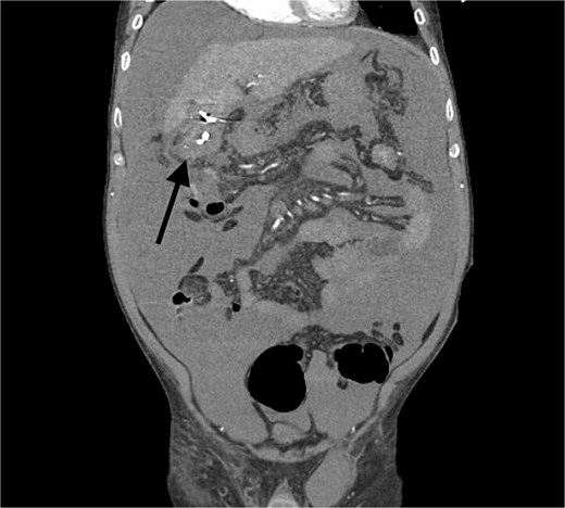
Pericholecystic hematoma without delineation of the inferior margins of the gallbladder.
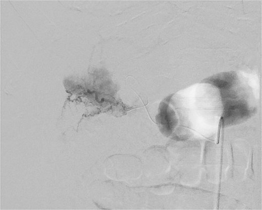
Small cystic artery pseudoaneurysm with complex perfusion.
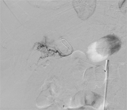
Post-embolization of cystic artery pseudoaneurysm.
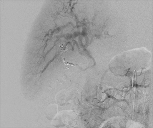
Post-embolization of cystic artery pseudoaneurysm.
The patient continued to require multiple transfusions despite embolization. The patient had a high-risk status with Model for End-Stage Liver Disease (MELD) score of 35. A transplant offer was expected with such a high MELD score. With the goal of stabilizing him before the transplant, the decision was made to proceed with open cholecystectomy after discussing surgical risks.
Intra-operatively, a midline incision with the right lateral extension was made. Two liters of ascites were drained. The gallbladder was found to be partially eviscerated from omental adhesions with an actively oozing anterior wall, which likely accounted for persistent bleeding. The gallbladder was dissected in a top-down fashion, with ligation and clipping of the cystic duct and artery. Hemostasis was achieved using electrocautery, argon beam coagulation, and topical hemostatic agents. The gallbladder and ascitic fluid were sent for pathology and cultures. Estimated blood loss was 125 ml, and the patient remained hemodynamically stable throughout.
During hospitalization for post-operative recovery, his MELD decreased to 23. A deceased donor liver became available, and the patient subsequently underwent orthotopic liver transplantation with concurrent primary umbilical hernia repair. He followed an appropriate postoperative recovery course after transplant. The pathology resulted for acute and chronic cholecystitis with cholelithiasis.
Discussion
Gallbladder hemorrhage is an exceptionally rare complication following colonoscopy and presents as a diagnostic challenge especially in advanced liver disease patients [4]. Less than 10 cases of cholecystitis after colonoscopy have been reported in the literature. Most reported patients presented with abdominal pain, anemia, or hemodynamic instability, and management ranged from supportive care to cholecystectomy [1]. This case is unique because it demonstrates a combination of gallbladder haemorrhage, cystic artery pseudoaneurysm, and intraoperative evidence of partial gallbladder avulsion in a patient with decompensated cirrhosis awaiting liver transplantation.
Hemorrhagic cholecystitis has been associated with trauma, anticoagulation, vascular abnormalities, and severe gallbladder wall inflammation, but its occurrence following colonoscopy is sporadic [4, 5]. The pathophysiology of gallbladder injury after colonoscopy remains poorly understood. Proposed mechanisms include dehydration-induced biliary stasis, increased intraluminal pressure, bowel distension, and direct mechanical trauma resulting from scope manipulation. In patients with pre-existing gallbladder disease, adhesions, chronic inflammation, or tissue friability, these forces may be amplified [2, 3]. We think pathophysiology in this case is multifactorial. Advanced cirrhosis and underlying coagulopathy likely increased susceptibility to haemorrhage, while mechanical forces generated during colonoscopy may have produced traction on the gallbladder and surrounding adhesions. Operative findings of partial avulsion of the gallbladder from omental adhesions with active bleeding from the anterior wall suggest a traumatic mechanism. Furthermore, subsequent development of a cystic artery pseudoaneurysm suggests vascular injury. The final pathology findings demonstrating acute and chronic cholecystitis, which may have further predisposed the gallbladder to injury.
Diagnosis of intra-abdominal pathology in cirrhosis patients is challenging. They present with absent classical signs such peritonitis, leukocytosis, or localized tenderness due to immune dysfunction, baseline ascites, and altered inflammatory responses [6]. Additionally, chronic anemia and coagulopathy may obscure the acuity of ongoing hemorrhage. Intraluminal blood can resemble sludge on imaging [7]. Clinicians evaluating abdominal pain after colonoscopy typically focus on more common complications such as colonic perforation, post-polypectomy bleeding, splenic injury, or mesenteric injury. However, gallbladder hemorrhage should be considered when patients develop unexplained anemia, recurrent hemorrhagic ascites, persistent abdominal pain, or transfusion requirements despite inconclusive imaging studies. In our patient, stable vitals and hemoglobin delayed definitive intervention, highlighting how gallbladder haemorrhage in cirrhosis can evolve insidiously rather than presenting as an overt surgical emergency.
Management of gallbladder hemorrhage requires an individualized, multidisciplinary approach. In hemodynamically stable patients, cross-sectional imaging and angiography can help identify vascular injuries and guide treatment. For cystic artery pseudoaneurysms, transarterial embolization is considered first-line therapy and can provide effective hemorrhage control [8]. However, embolization alone may be insufficient when structural gallbladder injury persists. Continued transfusion requirements, recurrent hemoperitoneum, ongoing hemorrhagic ascites, or persistent symptoms despite successful embolization should prompt consideration of operative intervention. In our patient, angiographic embolization successfully treated the pseudoaneurysm but failed to eliminate the underlying source of bleeding, as evidenced by ongoing transfusion dependence. Surgical exploration subsequently revealed a partially avulsed gallbladder with active bleeding that was not amenable to endovascular therapy alone.
An additional important aspect of this case is the role of cholecystectomy as a bridge to liver transplantation. Surgical planning in advanced liver disease requires careful assessment of risk versus benefits. Although cholecystectomy carries substantial morbidity in patients with advanced liver disease, ongoing hemorrhage represented a greater immediate threat to this patient than operative intervention. Successful haemorrhage control allowed stabilization of the patient’s condition, and he subsequently underwent orthotopic liver transplantation with favorable outcomes. To our knowledge, there is limited literature where successful surgical stabilization as a bridge to transplantation is highlighted. [9, 10]. This experience suggests that timely surgical intervention should not be deferred solely because of advanced liver disease when ongoing bleeding persists despite less invasive measures.
Conflicts of interest
None declared.
Funding
None declared.


