


Abstract
Gallstone ileus is a rare cause of mechanical bowel obstruction, predominantly affecting elderly patients with multiple comorbidities. We present a series of three patients illustrating the variability in clinical presentation, imaging findings, and surgical management. An 86-year-old male underwent successful laparoscopic enterolithotomy with an uneventful postoperative course and discharge on postoperative day 4. In contrast, two female patients (aged 77 and 84 years) required open surgery due to more severe clinical presentation and developed significant postoperative complications, including prolonged intensive care unit stay and delayed recovery. One case involved an atypical localization of the gallstone in the duodenum, representing a variant within the spectrum of Bouveret syndrome. These cases highlight the importance of early diagnosis and individualized surgical management. Minimally invasive approaches may offer improved postoperative outcomes in selected patients; however, careful patient selection remains crucial.
Introduction
Gallstone ileus is an uncommon but important cause of mechanical bowel obstruction, accounting for ~1%–4% of cases, with a higher prevalence in elderly patients [1, 2]. It typically develops as a complication of long-standing cholelithiasis, when inflammation leads to the formation of a cholecystoenteric fistula, allowing gallstones to enter the gastrointestinal tract [2].
Clinical presentation is often nonspecific, which may delay diagnosis. Computed tomography (CT) is considered the diagnostic modality of choice, particularly when features of Rigler’s triad are present [3, 4].
Surgical treatment remains the mainstay of management, most commonly in the form of enterolithotomy [5, 6]. However, the choice between open and minimally invasive approaches depends on the patient’s clinical condition and intraoperative findings.
Case reports
Case 1
An 86-year-old male with a history of diabetes mellitus and hypertension presented with a 2-day history of abdominal pain and long-standing intermittent biliary symptoms. Physical examination revealed mild abdominal distension without signs of peritonitis. Laboratory tests were within normal limits.
Contrast-enhanced CT demonstrated gallstone ileus with a cholecystoenteric fistula and an obstructing gallstone measuring 2.5 × 2.5 cm in the small intestine (Fig. 1).
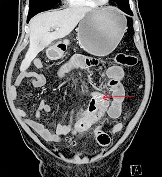
Contrast-enhanced CT demonstrating gallstone ileus with pneumobilia, bowel obstruction, and ectopic gallstone. The arrow indicates the ectopic gallstone.
The patient underwent laparoscopic enterolithotomy. Pneumoperitoneum was established using a Veress needle, and four trocars were inserted. Dilated jejunal loops and inflammatory adhesions were identified. The transition zone was located ~100 cm distal to the ligament of Treitz. A longitudinal enterotomy was performed proximal to the obstruction, and the gallstone was extracted and removed using an endoscopic retrieval bag (Fig. 2). The enterotomy was closed transversely in two layers.
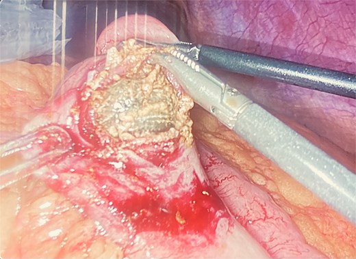
Laparoscopic extraction of gallstone through enterotomy.
The procedure lasted ~60 min without conversion. The postoperative course was uneventful, and the patient was discharged on postoperative day 4.
Case 2
A 77-year-old female with multiple comorbidities was admitted with hematemesis and vomiting. Gastroscopy did not reveal an active bleeding source. The clinical course was complicated by pneumonia and dehydration.
CT imaging demonstrated a cholecystoenteric fistula and findings consistent with Rigler’s triad, with a 3.5 × 3 cm gallstone located in the proximal ileum (Fig. 3).
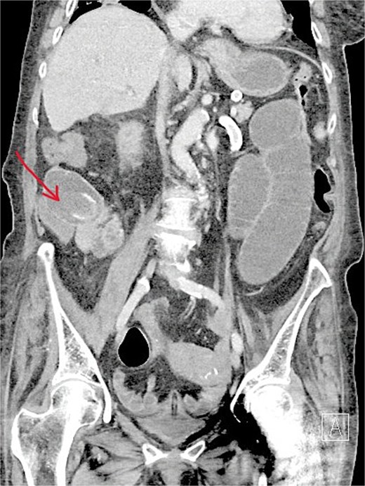
CT showing features of gallstone ileus with obstructing gallstone in the proximal ileum. The arrow indicates the ectopic gallstone.
An open enterolithotomy via laparotomy was performed. The transition point was identified ~150 cm proximal to the ileocecal valve. The stone was extracted through a longitudinal enterotomy, which was subsequently closed (Fig. 4).
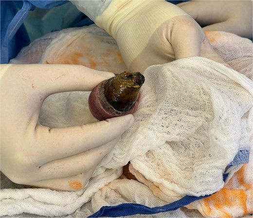
Open enterolithotomy with extraction of gallstone.
The postoperative course was complicated, requiring 10 days of intensive care. Reoperation for fascial dehiscence was necessary on postoperative day 5. The total hospital stay was 16 days.
Case 3
An 84-year-old female with known cholelithiasis was admitted with abdominal pain, vomiting, and collapse. CT imaging demonstrated gallstone ileus with a large gallstone (5.5 × 5 cm) impacted in the first part of the duodenum (Fig. 5). Prior imaging had demonstrated chronic cholecystitis without evidence of a cholecystoduodenal fistula (Fig. 6).
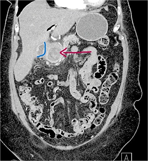
CT showing large gallstone impacted in the first part of the duodenum (D1). The arrow indicates the impacted gallstone.
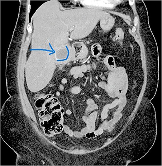
CT demonstrating chronic cholecystitis and cholelithiasis. The arrow indicates a gallstone within the gallbladder.
Gastroscopy with attempted stone removal was unsuccessful. The patient underwent open duodenolithotomy via laparotomy. The gallstone was extracted through a longitudinal duodenotomy, which was subsequently closed (Fig. 7).
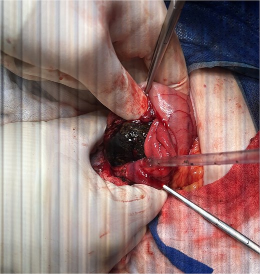
Duodenolithotomy with extraction of impacted gallstone.
The postoperative course was complicated by bronchopneumonia, atrial fibrillation, wound infection, and the development of a duodenocutaneous fistula, which was managed conservatively. The patient required 5 days of intensive care and was discharged after 12 days. The fistula persisted for two months postoperatively and resolved with conservative management.
Discussion
Gallstone ileus remains a diagnostically and therapeutically challenging condition, particularly due to its nonspecific presentation and the advanced age of affected patients. CT imaging plays a key role in diagnosis, with reported sensitivity of ~90%–93% and high specificity [4, 6]. The presence of Rigler’s triad significantly increases diagnostic confidence, although it is not present in all cases [3].
Our cases demonstrate the variability in presentation and outcomes. The patient treated laparoscopically had a favorable postoperative course, whereas patients requiring open surgery experienced more complicated recoveries [7, 8]. These differences are likely related to comorbidity burden and disease severity; however, they also suggest a potential benefit of minimally invasive surgery in appropriately selected patients [5, 9].
Laparoscopic enterolithotomy is technically demanding but may be a safe and effective option in selected patients [8, 10]. Careful patient selection remains essential. In our series, the only patient treated laparoscopically experienced the shortest hospital stay and an uncomplicated recovery, further supporting this approach in selected cases.
The third case represents an atypical presentation, overlapping with Bouveret syndrome, which may require alternative management strategies [11].
Conclusion
Gallstone ileus is a rare condition with variable clinical presentation. Minimally invasive surgery may offer improved postoperative outcomes in selected patients; however, individualized decision-making remains essential.
Conflicts of interest
The authors declare no conflict of interest.
Funding
No funding was received.
Consent statement
Written informed consent was obtained from the patients.


