


Abstract
Choledocholithiasis may still occur after cholecystectomy, whereas laparoscopic transcystic common bile duct exploration (LTCBDE) has rarely been reported in postcholecystectomy patients. A 58-year-old woman presented with a 1-day history of right upper quadrant pain 10 years after laparoscopic cholecystectomy. Magnetic resonance imaging and magnetic resonance cholangiopancreatography revealed a distal common bile duct stone of ~4 mm and common bile duct dilation to 12 mm. Intraoperatively, a cystic duct remnant measuring 0.8 cm in length and 4 mm in diameter was identified. A choledochoscope was inserted through the remnant into the common bile duct, and the stone was successfully removed. The patient recovered uneventfully and was discharged on postoperative Day 5. During 6 months of follow-up, no stone recurrence or biliary complications were observed. This case suggests that LTCBDE may be technically feasible in selected postcholecystectomy patients with a suitable cystic duct remnant and favorable biliary anatomy.
Introduction
Choledocholithiasis may still occur after cholecystectomy and can lead to obstructive jaundice, acute cholangitis, or biliary pancreatitis [1–4]. Although endoscopic retrograde cholangiopancreatography (ERCP) combined with endoscopic sphincterotomy is a commonly used treatment for common bile duct stones, it may still cause complications such as pancreatitis, bleeding, and perforation, and sphincterotomy may impair long-term sphincter of Oddi function [5–9]. Laparoscopic common bile duct exploration (LCBDE) provides an alternative one-stage approach and can be performed either through the cystic duct (LTCBDE) or through choledochotomy [10]. In patients with previous cholecystectomy, the absence of the cystic duct and postoperative anatomical changes often make the transcystic approach difficult to perform. We report a case in which LTCBDE was successfully performed through a preserved cystic duct remnant in a patient with previous cholecystectomy, highlighting the feasibility of this approach in carefully selected patients with favorable anatomy.
Case presentation
A 58-year-old woman was admitted to our hospital with a 1-day history of right upper quadrant pain. She had undergone laparoscopic cholecystectomy 10 years earlier and had a history of cerebral infarction 3 months earlier without obvious sequelae. Abdominal computed tomography (CT) performed in a local hospital showed choledocholithiasis. At the time of admission, physical examination revealed right upper quadrant tenderness without rebound pain or muscle tension. Laboratory tests showed inflammatory changes and abnormal liver function, including elevated bilirubin and liver enzyme levels, suggesting biliary obstruction with an associated inflammatory response. Abdominal CT showed that there is a surgical clip near the cystic duct remnant after cholecystectomy, dilated the duct of the bile duct, and stones in the duct (Fig. 1A and B). Further magnetic resonance imaging (MRI) and magnetic resonance cholangiopancreatography (MRCP) showed a stone in the distal common bile duct. The stone was about 4 mm in size and the common bile duct was dilated to about 12 mm (Fig. 1C and D).
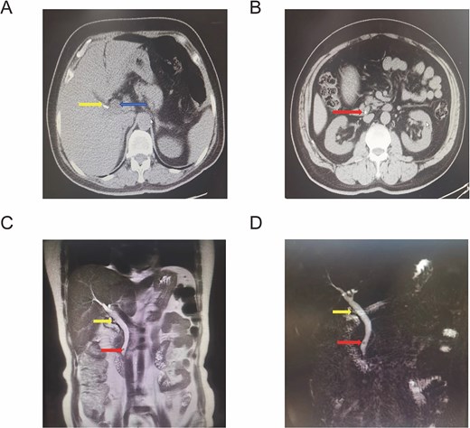
Preoperative imaging findings. (A) Abdominal CT showing clips at the cystic duct remnant after cholecystectomy and common bile duct dilatation. (B) Abdominal CT showing a common bile duct stone. (C) Coronal MRI showing the cystic duct remnant and a common bile duct stone. (D) MRCP showing the cystic duct remnant and a common bile duct stone.
The patient was diagnosed with choledocholithiasis. After consideration of the patient’s previous surgical history and preoperative imaging findings, laparoscopic common bile duct exploration was planned as a single-stage treatment option instead of ERCP. After preoperative preparation, laparoscopic exploration was performed under general anesthesia using a standard four-port technique with the patient in the supine position. Mild intra-abdominal adhesions were observed and it were carefully dissected to fully expose the hepatoduodenal ligament (Fig. 2A). The common bile duct was ~1.2 cm in diameter. A cystic duct remnant measuring ~0.8 cm in length and 4 mm in diameter was identified (Fig. 2B). The anterior wall of the cystic duct remnant was incised transversely ~1–2 mm proximal to its junction with the common bile duct (Fig. 2C). Bile outflow was then observed (Fig. 2D and E). After dilation of the cystic duct remnant, a 5-mm choledochoscope was introduced through the remnant (Fig. 2F and G). A black stone measuring ~4.5 mm in diameter was identified in the distal common bile duct and successfully extracted using a stone retrieval basket. The cystic duct remnant was closed with continuous sutures, and the stump was further reinforced with absorbable clips (Fig. 2H and I).
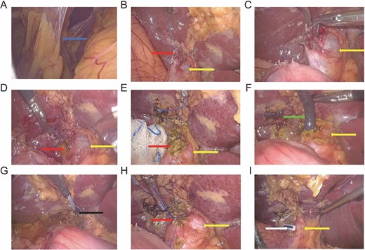
Intraoperative findings and surgical procedures of LTCBDE through the cystic duct remnant. (A) Intra-abdominal adhesions. (B) Dilatation of the common bile duct and the cystic duct remnant. (C) Transverse incision of the anterior wall of the cystic duct remnant ~1–2 mm proximal to its junction with the common bile duct. (D, E) Bile outflow after incision of the cystic duct remnant; the arrow indicates the cystic duct remnant. (F) Introduction of the choledochoscope through the cystic duct remnant; the yellow arrow indicates the common bile duct. (G) Extraction of the common bile duct stone. (H) Closure of the cystic duct remnant with sutures; the arrow indicates the common bile duct. (I) Closure of the cystic duct remnant with absorbable clips; the arrow indicates the common bile duct.
Postoperatively, the patient received anti-infective and supportive treatment. Oral intake was resumed on postoperative Day 3, and bilirubin levels and liver enzymes gradually returned to normal. The patient was discharged on postoperative Day 5. During a 6-month follow-up period, the patient recovered well without recurrence of stones or related complications.
Discussion
In this case, we successfully performed LTCBDE through a cystic duct remnant in a patient with previous cholecystectomy. This finding suggests that the transcystic approach may still be technically feasible in selected patients when a usable cystic duct remnant is present.
The successful use of the transcystic approach in this case was closely related to both preoperative evaluation and favorable anatomy. Preoperative imaging showed a dilated bile duct, a relatively small stone, and the presence of a cystic duct remnant, all of which supported selection of the transcystic approach. Intraoperative exploration further confirmed that the cystic duct remnant measured ~0.8 cm in length and 4 mm in diameter, which allowed insertion of the choledochoscope. In addition, the small stone size, limited number of stones, and patency of the distal bile duct also contributed to the successful completion of transcystic exploration. In contrast, in cases with a very short or narrow cystic duct remnant, severe intra-abdominal adhesions, large or multiple stones, or distal bile duct stenosis, the transcystic approach may be difficult to perform [10, 11].
In clinical practice, successful transcystic bile duct exploration also depends on operative technique. Careful identification of the cystic duct remnant is required, and clues such as sutures or clips left from previous surgery may help in locating the remnant. Adequate dissection of the remnant to its junction with the common bile duct and adjustment of the angle between the cystic duct and the common bile duct may facilitate the passage of the choledochoscope [12]. The remnant should be incised carefully to minimize injury to the bile duct wall, and dilation of the spiral valves of Heister may be performed when necessary to allow passage of the choledochoscope [13]. After stone removal, repeated exploration of the distal common bile duct should be performed to confirm complete clearance of stones. The need for drainage should then be determined according to the degree of biliary inflammation and the patency of the distal bile duct.
Several limitations of this report should also be acknowledged. This study describes a single case and therefore cannot determine the general applicability of this technique. The follow-up period was relatively short, and long-term outcomes such as stone recurrence or biliary stricture could not be fully evaluated. Further accumulation of cases and additional studies are needed to better define the indications and clinical value of this technique in patients with choledocholithiasis after cholecystectomy.
In conclusion, this case suggests that LTCBDE may still be feasible in patients after cholecystectomy when favorable anatomical conditions are present and adequate preoperative evaluation is performed. This approach may provide a minimally invasive and effective treatment option for selected patients, although the indications should be carefully considered.
Conflicts of interest
The authors declare that they have no conflict of interest.
Funding
This study was supported by the Guo Zhong Health Care Project of the China General Technology Group (GZKJ-KJXX-QTHT-20230626, 20240429).
Informed consent
Written informed consent was obtained from the patient for the publication of this case report and accompanying images.
Patient perspective
The patient was satisfied with the treatment outcome and the recovery process.
Approval of the research protocol
This study was conducted with approval from the Medical Ethics Committee of Beijing Electric Power Hospital, State Grid Corporation of China.


