


 https://orcid.org/0000-0003-0054-5652

Abstract
Amelia is a rare congenital malformation characterized by the complete absence of one or more limbs. We report the case of a 17-year-old boy with bilateral upper limb amelia and progressive thoracic scoliosis. He was fully independent in daily living, relying on his lower limbs for all activities. Radiographs demonstrated an 86° thoracic curve (T6–T12), skeletal maturity, and limited flexibility. Theoretical fusion to L3 or L4 risked major functional decline. He underwent selective thoracic fusion (T3–L1), sparing four lumbar discs. At 2-year follow-up, Cobb angle improved to 34°, SRS-22 score rose from 2.8 to 3.55, and independence was preserved. This case highlights the need to balance deformity correction with functional preservation in scoliosis management for patients with amelia.
Introduction
Amelia, defined as the congenital absence of one or more limbs, is a rare anomaly with an estimated incidence of 1.5 per 100 000 live births (Fig. 1) [1, 2]. It may occur in isolation or as part of multisystem syndromes and has been linked to both environmental and genetic etiologies. In bilateral upper limb amelia, scoliosis is almost universally observed, yet the medical literature contains little discussion of surgical correction strategies. This lack of guidance poses a challenge for spine surgeons tasked with balancing deformity correction against the preservation of function.
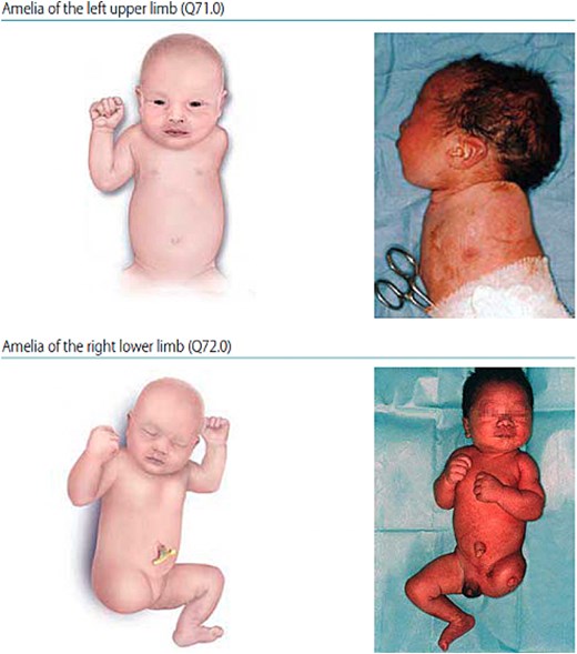
Clinical presentation of congenital bilateral upper limb amelia [1].
In adolescent idiopathic scoliosis (AIS), the determination of surgical strategy typically prioritizes coronal and sagittal correction, with cosmesis often a driving factor [3, 4]. For individuals with amelia, however, the situation is fundamentally different: upper limb absence necessitates complete reliance on the lower limbs for all daily living activities. Consequently, lumbar mobility is critical to independence, and a long fusion may carry devastating functional consequences. We present a case of scoliosis correction in bilateral upper limb amelia that demonstrates how surgical decision-making must be reframed in this rare and complex context.
Case report
A 17-year-old male with congenital bilateral upper limb amelia presented with progressive spinal scoliosis. He had adapted impressively to his disability, using his feet to feed, write, groom, and manipulate electronic devices. He lived independently with minimal assistance and was academically successful. However, in the preceding 2 years, he noted worsening truncal asymmetry, postural imbalance, and concern about potential progression of his scoliosis.
On physical examination, he had no upper extremities, a significant left thoracic rib prominence, and truncal shift. Lumbar flexibility was preserved and neurological examination was normal. His gait was steady and unaided. No cutaneous stigmata were present. Psychologically, he displayed resilience but emphasized that his priority was maintaining independence, even at the cost of cosmetic appearance.
Radiographs demonstrated an 86° left thoracic curve (T6–T12) with skeletal maturity (Risser 5, Sanders 7). The fulcrum unbending Cobb was 66° with only 23% flexibility, confirming a stiff curve. The Harrington stable zone was at L4 and the last touched vertebra was L3, indicating that conventional planning would have required fusion to at least L3. Magnetic resonance imaging excluded intraspinal anomalies (Fig. 2).
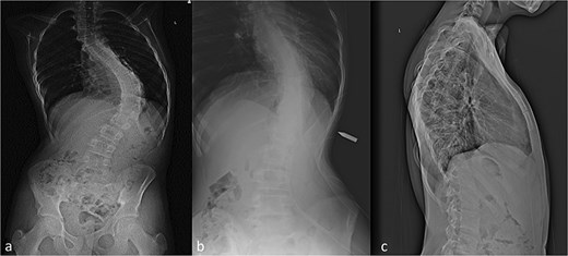
Preoperative whole spine radiographs. AP (a), bolster (b), and lateral (c) views showing 86° thoracic scoliosis with left convexity.
Several management options were discussed. Nonoperative management risked continued progression and potential loss of balance. Long posterior fusion from T3 to L3/L4 would likely achieve stable correction but would compromise lumbar mobility essential for his independence. Selective thoracic fusion (STF) offered a compromise, addressing the thoracic curve while sparing the lumbar spine. Anterior scoliosis correction and vertebral body tethering were considered inappropriate given rigidity and skeletal maturity.
The patient and family opted for STF. A posterior approach was used with pedicle screw instrumentation from T3 to L1. Correction was achieved using rod derotation and translation. Four lumbar discs were spared. Neuromonitoring signals remained stable throughout. Postoperative recovery was uneventful, with mobilization on Day 2 and discharge on Day 5.
At 2-year follow-up, the Cobb angle improved to 34°. Radiographs demonstrated stable correction without adding-on or coronal decompensation. The patient remained fully independent in all activities of daily living and reported improved posture. His SRS-22 score improved from 2.8 preoperatively to 3.55 postoperatively (Fig. 3).
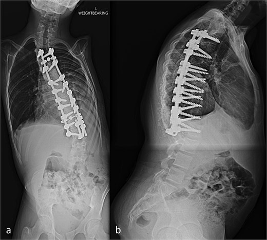
Postoperative whole spine radiographs. AP (a) and lateral (b) views showing improved alignment with Cobb angle reduced to 34° after selective thoracic fusion.
Discussion
This case illustrates the rare intersection of scoliosis and bilateral upper limb amelia. In typical AIS management, fusion levels are chosen based on stable vertebra alignment and risk of curve progression. In this patient, theoretical planning suggested fusion to L3 or L4. However, given his reliance on lumbar mobility, such an approach would have undermined his independence.
STF is a recognized strategy in AIS with nonstructural lumbar curves. While the lumbar curve in this case had structural elements, functional considerations outweighed strict radiographic indications. By limiting fusion to T3–L1, we were able to halt progression while preserving mobility critical to independence. Although risks of adding-on or coronal decompensation exist, these did not materialize at 2-year follow-up [5].
Alternative motion-preserving techniques such as vertebral body tethering were unsuitable due to curve stiffness and skeletal maturity. In younger patients with more flexible curves, such options might be considered. For this patient, however, posterior fusion was the only viable intervention [6, 7].
This case also highlights broader ethical considerations. For many adolescents with scoliosis, cosmetic outcome is a dominant concern. Here, the patient prioritized autonomy and independence. The decision-making process underscores the importance of tailoring surgical goals to patient-specific values.
The long-term outlook remains uncertain, with risks of adjacent segment degeneration and coronal imbalance requiring ongoing surveillance. Nonetheless, the positive 2-year outcome suggests that STF can be an appropriate compromise in this rare population. This report contributes to the limited body of literature and underscores the necessity of multidisciplinary planning that integrates surgical, rehabilitative, and psychosocial expertise.
Acknowledgements
The authors thank the patient and his family for their cooperation, and the multidisciplinary team for their involvement in care.
Conflicts of interest
The authors declare no conflicts of interest.
Funding
None declared.


