


 https://orcid.org/0000-0001-7843-0180

Abstract
Internal hernias are particularly difficult to diagnosis and should be considered in patients presenting with sudden, severe abdominal pain and symptoms of intestinal obstruction. Internal hernias carry a high risk of morbidity and mortality. Here, we present an unusual case of internal hernia through the foramen of Winslow of an ileoileal anastomosis created 25 years prior that was successfully diagnosed and operatively reduced via single-port laparoscopy with an optimal clinical outcome. Uniquely, the defect was closed using a ligamentum teres buttress to avoid undue tension at the foramen.
Introduction
Internal hernias are responsible for an estimated 0.2%–0.9% of intestinal obstructions [1]. Herniation through the epiploic foramen of Winslow (FoW), known by the eponym of Blandin’s hernia, is rarer still, accounting for ~8% of internal hernias [2]. The organs typically implicated in foramen of Winslow hernias (FWH) include small bowel (63%), cecum and right colon (30%), and transverse colon (7%) [2]. The challenge of timely diagnosis leads to many patients presenting with strangulated bowel from closed-loop obstruction, resulting in high mortality rates, up to 49%. Surgical closure of the FoW to prevent recurrence is controversial. Given its rarity, the body of literature describing the diagnosis and management for this condition is limited to case reports and reviews of case series. Multiple etiologies for this condition have been proposed, including changes in intra-abdominal pressure, abnormal enlargement of the FoW and excessive visceral mobility (i.e. elongated mesentery) [2]. Operative intervention is the preferred treatment modality for this condition. The decision for exploratory laparotomy versus initial diagnostic laparoscopy is driven by the patient’s hemodynamic stability, available surgical resources, and the suspicion of bowel strangulation/ischemia preoperatively. The primary controversy in operative management is the decision to resect the herniated organs, perform a pexy procedure, or suture close the foramen to prevent recurrence, which has yet to be reported in the literature.
We present the unique case of a patient with a remote history of total abdominal colectomy and ileal pouch-anal anastomosis that developed ileoileal anastomosis incarceration through the FoW which was managed with minimally invasive techniques and a novel means to perform a tension-free obliteration of the foramen.
Case report
The patient is a 55-year-old male with a history of right partial nephrectomy for renal cell carcinoma and ulcerative colitis requiring multiple operations, including a total abdominal colectomy (TAC) with end ileostomy, ileal pouch-anal anastomosis (IPAA) with diverting loop ileostomy (DLI), and subsequent ileostomy reversal nearly 25 years ago. He presented with twelve hours of sudden-onset, severe epigastric pain, and nausea. The day prior, he was passing flatus with non-bloody bowel movements. His heart rate on arrival was 100 beats-per-minute; the remainder of his vitals were unremarkable. Physical examination was notable for mild distension and focal epigastric tenderness without peritonitis. Laboratory analysis showed mild leukocytosis of 10.8 k/μL and lactic acidosis of 5.1 mmol/L. Computed tomography (CT) abdomen-pelvis with intravenous contrast showed 7.4 cm of distended small bowel that passed between the inferior vena cava and left hepatic lobe, resting just cephalad to the stomach suggesting an FWH (Fig. 1). Decompressed small bowel loops passed posteriorly to the portal triad, consistent with closed-loop obstruction. The portal vein appeared mildly narrowed and ‘stretched.’ There was no evidence of ischemia or perforation. The patient was taken urgently to the operating room for diagnostic laparoscopy.
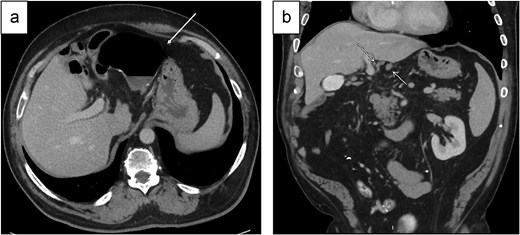
(a) Axial and (b) coronal views of the ileoileal anastomosis herniating through the FoW and resting in the lesser sac cephalad to the stomach. Arrows indicate the (a) herniated bowel and (b) proximal and distal segments of collapsed ileum passing posterior to the portal triad consistent with closed-loop obstruction.
Single-port laparoscopy was established via Hasson technique. The ileoileal anastomosis was protruding into the lesser sac and was visible through the pars flaccida of the gastrohepatic ligament (Fig. 2a). In a lateral-to-medial orientation, two loops of small bowel entered the FoW over the retroperitoneum (Fig. 2b). The herniated ileum was reduced with careful enterolysis and inferolateral traction (Fig. 3). There was no threatened or ischemic bowel. The FoW appeared patulous and not amenable to direct closure. The natural position of the reduced ileum, directly adjacent to the foramen, was worrisome for potential re-herniation. The ligamentum teres hepatis was mobilized and sutured to the retroperitoneal fat and anterosuperior duodenal serosa using 0 silk suture, occluding the orifice. The abdomen was subsequently de-sufflated under direct visualization and closed.
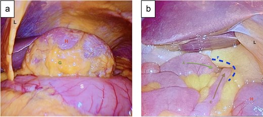
(a) Intraoperative image of the ileoileal anastomosis protruding through the FoW into the lesser sac and resting just cephalad to the stomach. (b) Two limbs of small bowel entering the FoW laterally. G: gastrohepatic ligament; H: herniated bowel; L: ligamentum teres hepatis; A: afferent limb; E: efferent limb; F: FoW; S: stomach.
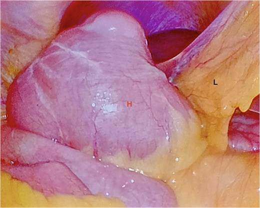
Dilated but viable ileoileal anastomosis after reduction from the FoW. H: herniated bowel (now reduced); L: Ligamentum teres hepatis.
The patient recovered quickly, passed flatus, and had the nasogastric tube removed on postoperative day 1. He advanced from clear liquids to a regular diet without issue and was discharged home on postoperative day 2. He is doing well with no suspicion of recurrence 1.5 years postoperatively.
Discussion
Internal herniation through the FoW is an exceedingly rare clinical entity associated with a high mortality rate from delay in diagnosis and bowel strangulation. We present an unusual case of ileoileal anastomosis FWH in patient with a complex abdominal surgical history. The patient was diagnosed after recognition of a closed-loop bowel obstruction at the FoW. Though subtle, the patient’s narrowed portal vein on imaging supported the FWH diagnosis. He subsequently underwent successful reduction with single-port laparoscopy and obliteration of the FoW with a novel, tension-free technique using a pedicle of the ligamentum hepatis with salutary results.
One theory for FWH formation is excessive mesentery length [2]. While no lengthening was observed at the herniation site, it is possible that small bowel and mesentery folding from the anti-peristaltic side-to-side, functional end-to-end anastomosis provided a lead point, promoting herniation. Partial right nephrectomy and TAC with mobilization of the ileum for both an IPAA and reversal of a DLI results in loss of natural anatomic barriers that may prevent such a hernia from occurring.
FoW herniation after colon resection was described once in the literature. An iatrogenic defect in the lesser sac, created during a laparoscopic subtotal colectomy, permitted medial-to-lateral small bowel FHW, resulting in strangulation 15 days postoperatively [3]. Both the lesser sac and foramen were sutured closed after a bowel resection was performed. Similarly, we elected to close the FoW. However, we used a ligamentum teres hepatis pedicle to obliterate the foramen without tension given anatomic constraints of direct suture closure. On review of the case report literature on laparoscopic management of FWH, the decision to close the FoW appears to be largely surgeon preference [2], as there have not been descriptions of FWH recurrence. Closure of the foramen, if performed, must be approached cautiously as injury to the portal vein and common bile duct is possible.
In summary, early diagnosis and reduction of FWH is crucial to prevent complications including bowel strangulation and necrosis. Loss of anatomical barriers, such as following colon resection, may permit FWH development. In patients where suture close would result in excessive tension of the FoW, a pedicled ligamentum teres may be utilized.
Conflicts of interest
The authors have no conflicts of interest pertinent to the contents of this original manuscript.
Funding
No funding source.
Ethics approval statement
Institutional Review Board review was not necessary for this work.


