


 https://orcid.org/0009-0000-4082-4838

Abstract
A pancreatic combined Grade 1 neuroendocrine tumour (NET) and ductal adenocarcinoma (PDAC) — meeting morphological criteria for both mixed neuroendocrine–non-neuroendocrine neoplasm (MiNEN) and collision tumour — is exceptionally rare. We report a 68-year-old female with multifocal pancreatic cystic lesions, main pancreatic duct dilatation, and elevated CA 19–9, with imaging features consistent with intraductal papillary mucinous neoplasm. Following multidisciplinary review, total pancreatectomy with duodenectomy and Roux-en-Y reconstruction was performed. Histopathology revealed PDAC (65%) and a well-differentiated Grade 1 PanNET (35%), with the latter confirmed by diffuse positivity for Synaptophysin and Chromogranin A and Ki-67 < 1%. A co-existing simple mucinous cyst with high-grade dysplasia was also identified. Adjuvant XELOX chemotherapy was administered after the patient declined infusional FOLFOX. This case highlights the diagnostic challenge of distinguishing MiNEN from collision tumour in the absence of molecular clonality data, and adds to the sparse literature on combined pancreatic NET–PDAC neoplasms.
Introduction
Mixed neuroendocrine–non-neuroendocrine neoplasms (MiNENs) are rare tumours defined by the simultaneous presence of a neuroendocrine and a non-neuroendocrine component, each constituting ≥30% of the tumour mass, per the 2019 WHO classification [1]. In the vast majority of pancreatic MiNENs the neuroendocrine component is a high-grade neuroendocrine carcinoma; the combination of a well-differentiated Grade 1 neuroendocrine tumour (NET) with ductal adenocarcinoma (PDAC) is exceptionally uncommon and raises a critical differential with collision tumour, in which two clonally unrelated neoplasms occur in close proximity [1, 2].
Intraductal papillary mucinous neoplasms (IPMNs) are the most frequent pancreatic cystic neoplasms and carry a significant risk of malignant transformation; main-duct type harbours high-grade dysplasia or invasive carcinoma in up to 70% of resected specimens [3]. The co-existence of multifocal IPMN with a concurrent combined pancreatic NET–PDAC neoplasm is extraordinarily rare [4, 5]. We present one such case and discuss the diagnostic dilemma between MiNEN and collision tumour, the rationale for total pancreatectomy, and the adjuvant chemotherapy decision-making process.
Case report
A 68-year-old female with hypertension, type 2 diabetes mellitus, and prior cholecystectomy presented with a several-month history of dyspepsia, bloating, altered bowel habits, and progressively worsening intermittent abdominal pain. Physical examination revealed only mild epigastric tenderness. Routine laboratory investigations were unremarkable except for an elevated serum CA 19–9 at 168.39 U/ml.
Cross-sectional imaging (contrast-enhanced CT and MRI) demonstrated an ill-defined intraductal lesion at the pancreatic head–body junction with main pancreatic duct (MPD) dilatation (~6 mm), and clustered cystic lesions involving the head, body–neck junction, and uncinate process communicating with the MPD. A solid enhancing mural nodule was identified within the dilated duct. Mild upstream biliary dilatation was also noted. Findings were most consistent with mixed-type IPMN (Fig. 1).
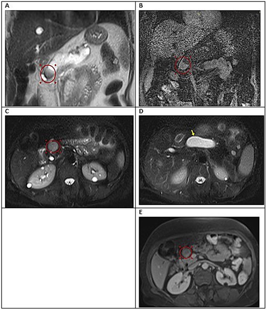
Dynamic MRI of the abdomen demonstrating features of mixed-type intraductal papillary mucinous neoplasm (IPMN). (A) Coronal T2-weighted image. (B) Axial post-contrast T1-weighted image. (C, D) Axial T2-weighted images. (E) Axial post-contrast T1-weighted fat-suppressed image in the portal venous phase. Across sequences, there is cystic dilatation of the main pancreatic duct along the body and tail (arrow), with a faintly enhancing intraductal focal lesion identified at the level of the pancreatic neck (circle).
Endoscopic ultrasound (EUS) confirmed cystic lesions at the pancreatic head and uncinate process, with a second lesion at the pancreatic neck communicating with the MPD. EUS-guided fine-needle biopsy yielded haemorrhagic and necrotic material with rare atypical cells of undetermined significance. Pancreatic cyst fluid CEA was elevated at >200 ng/dl, suggestive of a mucinous neoplasm with malignant potential.
The multidisciplinary team (MDT) recommended surgical resection despite non-specific cytology, on the basis of multiple high-risk features: a solid enhancing mural nodule, multifocal cystic disease (a 2.5 × 7 cm cyst), MPD diameter 6 mm, CA 19–9 of 168 U/ml, and cyst-fluid CEA >200 ng/dl. Total pancreatectomy — rather than pancreaticoduodenectomy or distal pancreatectomy — was chosen because the multifocal disease involved both pancreatic head and tail, with loss of normal parenchymal architecture replaced by an expansile dilated midline cystic structure (≈2.5 × 7.5 cm) coursing along the entire pancreatic duct. Any lesser resection would have carried a high risk of residual malignancy and the morbidity of a second pancreatic operation. Total pancreatectomy with duodenectomy and Roux-en-Y hepaticojejunostomy reconstruction was performed without complications.
Histopathology demonstrated PDAC (65%) and a well-differentiated PanNET Grade 1 (35%), with the two components appearing morphologically distinct rather than intermingled (Figs 2 and 3). The neuroendocrine component showed diffuse strong cytoplasmic positivity for synaptophysin and chromogranin A; the ductal component was negative for both. Ki-67 in the neuroendocrine component was <1%, supporting Grade 1 classification. A co-existing simple mucinous cyst with high-grade dysplasia was also identified. Postoperative imaging showed expected post-pancreatectomy changes with no acute complications.
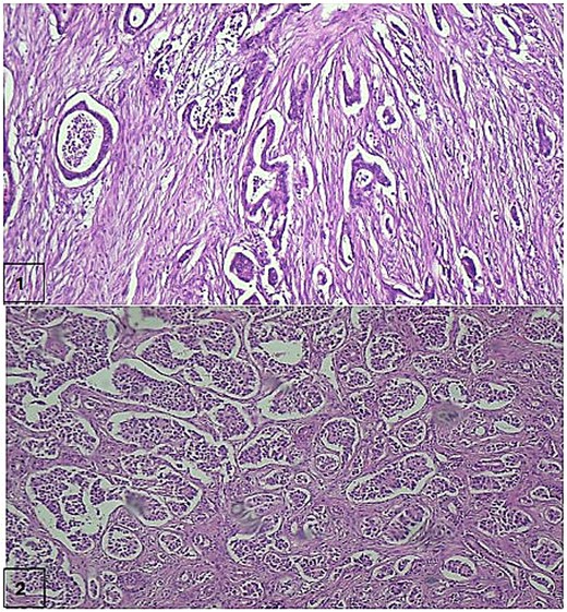
Histopathological features of the combined pancreatic G1 NET and PDAC. (1) The ductal adenocarcinoma component, composed of well-formed tubules lined by atypical epithelial cells embedded within desmoplastic stroma (H&E, ×10). (2) The neuroendocrine tumour component, comprising nests and trabeculae of small, monomorphic cells with scant eosinophilic cytoplasm and round nuclei with stippled chromatin (H&E, ×10).
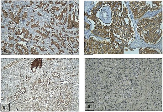
Immunohistochemical characterization of the combined neoplasm. (3) The neuroendocrine component demonstrates diffuse and strong cytoplasmic positivity for Synaptophysin (IHC, Synaptophysin, ×10). (4) The neuroendocrine component is also strongly positive for chromogranin A (IHC, chromogranin A, ×40). (5) The ductal adenocarcinoma component is negative for both Synaptophysin and chromogranin A (IHC, Synaptophysin, ×10). (6) The Ki-67 proliferation index in the neuroendocrine component is less than 1%, supporting classification as Grade 1 NET (IHC, Ki-67, ×10).
Adjuvant chemotherapy was initially planned as FOLFOX, but the patient declined the 46-hour continuous 5-FU infusion. As capecitabine is an oral fluoropyrimidine prodrug pharmacologically equivalent to infusional 5-FU, XELOX (Oxaliplatin 85 mg/m2 Day 1 plus Capecitabine 1000 mg/m2 twice daily on Days 1–14, every 21 days for four cycles) was selected as the oral equivalent of FOLFOX [6, 7].
Discussion
The principal teaching point of this case is the diagnostic challenge of classifying a pancreatic neoplasm composed of PDAC and a well-differentiated Grade 1 NET. By WHO 2019 criteria, MiNEN requires each component to constitute ≥30% of the tumour, with either intermingled or distinct borders [1]. Our case fulfils this threshold; however, the neuroendocrine component of MiNEN is, in the overwhelming majority of cases, a high-grade neuroendocrine carcinoma. The presence of a well-differentiated G1 NET is biologically unusual and raises a serious differential with collision tumour [2, 8].
Two features in our case favour consideration of a collision tumour: the G1 morphology of the neuroendocrine component, and the largely distinct (non-intermingled) borders between the two components. The WHO classification notes that in such combinations, the likelihood of a collision tumour is increased [1]. Definitive distinction requires molecular clonality analysis demonstrating whether the two components share a common origin — a study not available at our institution. We therefore present this case transparently as a combined G1 NET and PDAC of uncertain classification (MiNEN versus collision tumour), and recommend that future analogous cases ideally undergo molecular confirmation.
The MDT decision to proceed with resection despite non-diagnostic cytology was driven by the convergence of a solid enhancing intraductal mural nodule, multifocal main-duct involvement, MPD of 6 mm, CA 19–9 of 168 U/ml, and cyst-fluid CEA >200 ng/dl — together fulfilling high-risk stigmata for surgical management of pancreatic cystic neoplasms [3, 9]. Total pancreatectomy, although carrying long-term metabolic sequelae [10], was necessitated by the anatomical extent of disease, which precluded any lesser resection from achieving adequate margins.
No evidence-based consensus exists for adjuvant therapy in combined pancreatic NET–PDAC neoplasms. Guided by the dominant PDAC component, FOLFOX — an oxaliplatin-based regimen supported by Cochrane-level evidence [6] — was initially planned. Following the patient's informed refusal of the 46-hour 5-FU infusion, XELOX was selected as the oral fluoropyrimidine equivalent, an approach supported by published data on oxaliplatin-fluoropyrimidine combinations in PDAC [7, 11].
Our case has important limitations. First, the absence of molecular clonality studies precludes definitive distinction between MiNEN and collision tumour. Second, follow-up duration is limited, and long-term oncological outcomes for this combination remain unknown. Despite these limitations, this case adds to the sparse literature on combined pancreatic G1 NET and PDAC, and underscores the importance of multidisciplinary judgement when imaging, biochemical, and cytological findings do not align with a single pathological diagnosis.
Acknowledgements
The authors would like to thank the multidisciplinary team members, nursing staff, and the patient for permitting publication of this case.
Conflicts of interest
None declared.
Funding
This work received no specific funding from any funding agency in the public, commercial, or not-for-profit sectors.
Ethical approval
Ethical approval was not required for this single case report. The case was managed in accordance with institutional standards at Warith International Cancer Institute, Karbala, Iraq.
Consent
Written informed consent was obtained from the patient for publication of this case report and any accompanying images.
Guarantor
Ali Naser Aldarawsha.


