


Abstract
Comamonas testosteroni is an uncommon environmental Gram-negative bacillus rarely implicated in human infections. We report a case of a 34-year-old male who presented with acute appendicitis complicated by a sealed perforation. Laparoscopic appendectomy was performed, and postoperative cultures identified C. testosteroni in the appendiceal tissue, alongside Pseudomonas aeruginosa and Streptococcus anginosus in the peritoneal fluid. Initial empirical antibiotics were escalated to piperacillin–tazobactam based on culture results. The patient recovered uneventfully and was discharged on postoperative day nine. This case highlights the emerging clinical relevance of C. testosteroni and the importance of microbiological evaluation in guiding management of perforated appendicitis.
Introduction
Acute appendicitis is the most common surgical emergency globally. The bacteria most commonly isolated in association with appendicitis are Escherichia coli and Bacteroides fragilis [1]. However, An increasing number of reports have highlighted the involvement of Comamonas testosteroni, an aerobic, Gram-negative, motile bacillus, in intra-abdominal infections. This organism, the most frequently isolated species of the Comamonas genus, is uniquely named for its ability to utilize testosterone as its sole carbon source in vitro. Although historically considered to be of low virulence and rarely pathogenic to humans [2]. C. testosteroni has been increasingly associated with gastrointestinal infections, particularly appendiceal perforation complicated by peritonitis and bacteremia [3]. To date, nearly 52 cases implicating C. testosteroni in human disease have been reported [4]. This case report presents a rare instance of perforated appendicitis due to C. testosteroni, highlighting diagnostic challenges, microbiological findings, and postoperative management considerations.
Case presentation
A 34-year-old male with no significant past medical or surgical history presented to the Emergency Department with a one-day history of epigastric pain that migrated to the right lower quadrant. The pain was continuous, progressive, and dull in character, exacerbated by movement, and associated with anorexia, nausea, and a single episode of vomiting. Review of systems was otherwise unremarkable. On examination, the patient was febrile (38.6°C) and tachycardic (heart rate: 123 bpm). Abdominal examination revealed a soft, lax abdomen with marked tenderness and rebound tenderness localized to the right iliac fossa. Laboratory investigations demonstrated leukocytosis with a white blood cell (WBC) count of 12.2 × 10⁹/L, while hemoglobin and coagulation profiles were within normal limits.
A contrast-enhanced computed tomography (CT) scan of the abdomen revealed an enlarged appendix measuring 1.5 cm in diameter, with an enhancing wall, significant surrounding fat stranding, and intraluminal calcifications, consistent with acute appendicitis (Fig. 1). The patient underwent emergent laparoscopic appendectomy. Intraoperatively, the appendix was inflamed with a sealed perforation and localized contamination; a peritoneal drain was placed surrounding localized contamination; a peritoneal drain was placed.
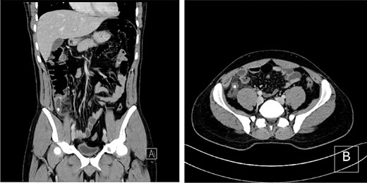
CT scan showing an inflamed, dilated appendix with surrounding fat stranding and intraluminal calcifications in (A) coronal and (B) axial views.
Postoperatively, the patient was initiated on intravenous ceftriaxone (2 g daily) and metronidazole (500 mg every 8 h). On postoperative day five, cultures from the resected appendix grew C. testosteroni, while peritoneal fluid cultures grew Pseudomonas aeruginosa and Streptococcus anginosus. Following infectious disease consultation, antibiotics were escalated to intravenous piperacillin–tazobactam (4.5 g every 6 h). After 4 days of targeted therapy, the patient reported resolution of abdominal pain and normalization of WBC count (5.7 × 10⁹/L). The drain was removed, and the patient was discharged with oral analgesics and outpatient follow-up. At the 2-month follow up, he remained asymptomatic and clinically stable.
Discussion
The genus Comamonas, within the family Comamonadaceae, was established in 1985 with C. terrigena as its initial species. In 1987, Pseudomonas testosteroni and Pseudomonas acidovorans were reclassified into this genus [2]. Currently, at least 17 species have been identified, including C. aquatica, Comamonas kerstersii, and C. denitrificans [3]. These are aerobic, Gram-negative, oxidase-positive, motile bacilli that grow on standard media and exhibit pink pigmentation [2]. The first human infection by C. testosteroni was reported in 1975 by Atkinson et al. [5] An earlier literature review of 41 reported cases found that intra-abdominal infections (18 cases) and bacteremia (15 cases) were among the most frequently encountered clinical presentations. Less frequent sites include the eye, urinary tract, endocardium, and central nervous system [3, 6]. C. testosteroni’s pathogenesis often involves translocation from the gastrointestinal tract, especially in the context of appendiceal perforation [3, 7].
Building on previous findings, we present the case of a 34-year-old male with no prior medical history who developed acute appendicitis complicated by a sealed perforation. Postoperative cultures identified C. testosteroni in the appendiceal tissue, and P. aeruginosa along with S. anginosus in the peritoneal fluid. This polymicrobial pattern aligns with earlier reports, including those by Barbaro et al. and others, in which C. testosteroni was isolated from polymicrobial infections following perforated appendicitis [2, 6]. Gul et al. similarly documented C. testosteroni bacteremia in a comparable clinical setting, further supporting the association between appendiceal perforation and systemic dissemination [8].
Initial empirical treatment with intravenous ceftriaxone and metronidazole was escalated to piperacillin–tazobactam following the identification of C. testosteroni in appendiceal tissue and P. aeruginosa and S. anginosus in peritoneal fluid. The patient demonstrated rapid clinical improvement and was discharged without complications. Previous literature has reported that C. testosteroni is generally susceptible to β-lactam/β-lactamase inhibitors, cephalosporins, carbapenems, and aminoglycosides [2, 3, 8]. Susceptibility to fluoroquinolones and trimethoprim–sulfamethoxazole has also been documented in multiple cases [2, 8], although resistance to both agents has also been noted [3]. Suggesting possible strain-specific variability. Compared to other published cases, such as bacteremia reported by Gul et al. [8], prolonged antibiotic therapy in Miloudi et al.’s case [3], and limited follow-up in Bayhan et al.’s report [2], our patient’s favorable outcome underscores the importance of timely surgical intervention and appropriate empirical therapy.
Conclusion
Comamonas testosteroni is a rare but emerging pathogen in intra-abdominal infections, particularly after appendiceal perforation. This case highlights the importance of thorough microbiological investigation and culture-directed antibiotic therapy. Early surgical intervention, combined with targeted antimicrobial treatment, can lead to favorable outcomes. Awareness of such atypical pathogens and their resistance profiles is essential for optimal management of complicated appendicitis.
Conflict of interest statement
None declared.
Funding
None declared.
