


Abstract
Vascular injuries can initially present with hemorrhage or limb-threatening conditions, and may later lead to complications such as pseudoaneurysms or arteriovenous fistulas. Pseudoaneurysms are a known complication of arterial injuries resulting from trauma. Proper management following trauma is crucial in preventing pseudoaneurysm development. Here we report a case of pseudoaneurysm arising from an unnamed branch of posterior tibial artery eroding bone in a 4-year old boy who presented with a non-pulsatile, non-tender swelling on the medial aspect of left leg. Early identification of these complications is crucial for preserving the limb, as demonstrated by the patient's uneventful postoperative recovery.
Introduction
Pseudoaneurysms are false aneurysms that develop at arterial damage sites caused by trauma or infection. Unlike true aneurysms, where the vessel wall bulges out, a pseudoaneurysm does not involve the vascular wall. Blood escapes from the injury site and is contained by a wall formed from the clotting cascade [1]. The most common pseudoaneurysms involve the heart, femoral artery, visceral organs, and aorta. Peripheral aneurysms of the lower extremities are rare in adults, typically affecting the popliteal artery (70%) and femoral artery (20%). Only ⁓10% occur in other regions [2]. Lower extremity pseudoaneurysms are uncommon but recognized, and their occurrence in children is particularly rare, with few reported cases. A total of 28 foot pseudoaneurysms have been documented in English literature based on a PubMed search. Among these, eight cases involved the distal posterior tibial artery, with six involving children [3, 4]. This report presents the first case of a post-traumatic pseudoaneurysm from an unnamed branch of the posterior tibial artery in a 4-year-old boy. This case highlights the importance of understanding pseudoaneurysm etiology and initial management to prevent their development.
Case report
A 4-year-old boy presented with swelling on the medial aspect of his left leg. Three months prior, the patient had sustained a left tibial shaft fracture, initially treated conservatively. The swelling developed gradually with localized tenderness. On examination, the swelling was firm and tender to palpation, with intact overlying skin (Fig. 1). Radiography revealed erosion of the tibia (Fig. 2). Given the patient's fracture history, the initial differential included osteomyelitis, and empirical antibiotics were started. The vascular surgery team suspected a pseudoaneurysm and recommended further evaluation. Doppler ultrasound confirmed a pseudoaneurysm, revealing an abnormal vascular structure with turbulent blood flow. To delineate the vascular anatomy and identify the pseudoaneurysm's origin, magnetic resonance angiography (MRA) was ordered. It demonstrated the pseudoaneurysm arising from an unnamed posterior tibial artery branch, likely injured during initial trauma. The aneurysmal sac was located near the fracture site, eroding the underlying tibial bone (Fig. 3). The pseudoaneurysm's size warranted surgical intervention. The patient underwent excision of the aneurysmal sac and vessel wall repair (Fig. 4). Under general anesthesia, the aneurysmal sac was dissected out (Fig. 5), and the posterior tibial artery wall was repaired with lateral sutures. Hemostasis was achieved, and the wound was closed in layers. The patient had an uneventful postoperative recovery.
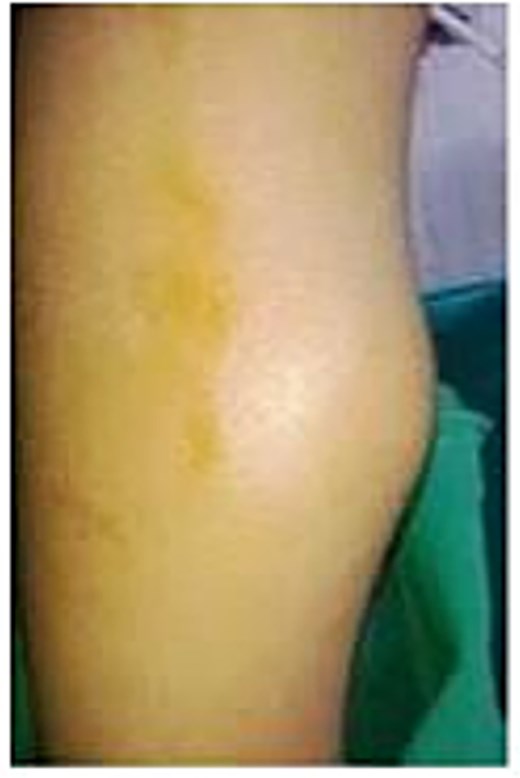
Clinical picture of the patient's left leg showing swelling on the medial aspect.
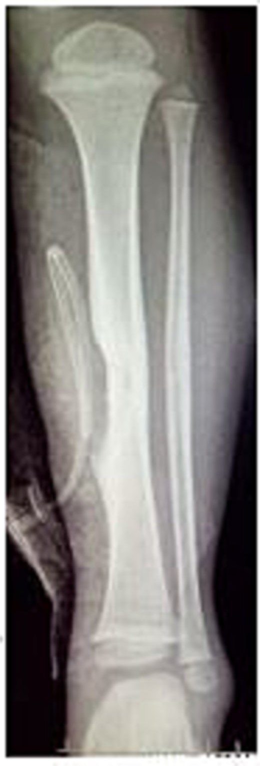
Radiograph showing erosion of tibia beneath pseudoaneurysm.
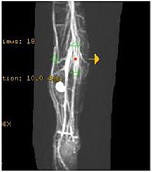
MRA showing the pseudoaneurysm arising from an unnamed branch of the posterior tibial artery.
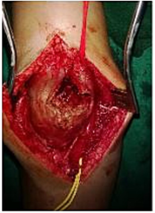
Intraoperative view of the surgical field.
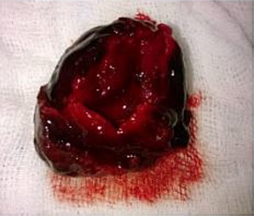
Excised pseudoaneurysm sac.
Discussion
Pseudoaneurysm of the posterior tibial artery is rare and typically results from trauma or iatrogenic complications [5–7]. Infections and autoimmune disorders can contribute to its development [5, 8]. In our case, trauma caused the pseudoaneurysm. Arterial aneurysms are uncommon in children, with fewer than 250 cases reported, and lower extremity cases are rarer, with fewer than 25 documented [3, 4, 9]. A pseudoaneurysm occurs when the arterial wall is compromised due to iatrogenic factors, trauma, or infection [10]. In our case, the arterial wall injury resulted from blunt trauma associated with the fracture. Unlike true aneurysms, pseudoaneurysms' walls consist of surrounding tissues with fibrin and platelets formed through clotting. While pseudoaneurysms typically occur in peripheral arteries after endovascular procedures, they can develop in any artery [1, 10]. In our case, the pseudoaneurysm developed in the posterior tibial artery following a tibial shaft fracture 3 months earlier.
The first reported arteriovenous aneurysm involving posterior tibialis vessels was described by Higgs after foot stabilization [11]. Morris and Morse later reported the first foot pseudoaneurysm affecting only the posterior tibialis artery [12]. Trauma, iatrogenic factors, connective tissue disorders, and infections commonly cause pseudoaneurysms [3]. In our patient, trauma from a left tibial shaft fracture was treated conservatively. Proper management following trauma is crucial in preventing pseudoaneurysm development [3]. Conservative treatment of the tibial fracture might have contributed to pseudoaneurysm formation. Primary care physicians should be vigilant about pseudoaneurysms in the medial leg following trauma, as early vessel repair could prevent the condition [3]. In children, pseudoaneurysms typically present as painful, pulsatile masses following trauma [4]. However, our patient had a non-pulsatile, non-tender swelling on the medial aspect of the left leg. In severe cases, patients may experience limb ischemia [4], which was not observed here. Pseudoaneurysms most often occur in the anterior tibial artery [13]. A posterior tibial artery pseudoaneurysm is rare and has been documented few times in literature [3–5, 9, 10, 14, 15]. The posterior tibial artery's anatomical protection makes it less vulnerable to injury [10]. Only two cases of posterior tibial artery pseudoaneurysm in the shank region have been reported in adults in the past decade [5, 10].
Ultrasound is a key diagnostic tool for aneurysms [16]. It is non-invasive, cost-effective, and requires no contrast or sedation in pediatric patients [17]. Ultrasonography can accurately locate posterior tibial artery aneurysms and detect mural thrombi [18]. In our case, Doppler ultrasound confirmed the pseudoaneurysm and helped assess perfusion for perioperative decisions. MRA and computed tomography angiography are alternative imaging options [4, 17]. Our MRA showed the pseudoaneurysm near the fracture site, eroding the tibial bone, originating from a posterior tibial artery branch. Treatment options include arterial repair, graft interposition, ligation, compression, thrombin injection, and endovascular intervention [6]. With no standard protocol for post-traumatic posterior tibial artery pseudoaneurysms, surgical repair is preferred over conservative treatment due to embolization risks [10]. The patient underwent aneurysmal sac excision and vessel wall repair.
Conclusions
Pseudoaneurysms are a known complication of arterial injuries resulting from trauma. Following trauma in children, arterial injury must be considered and promptly addressed with primary repair. While many cases present with a pulsatile mass, it is crucial to recognize that pseudoaneurysms can manifest as non-pulsatile swelling following limb trauma. Performing fine needle aspiration cytology on such swellings without proper preparation can lead to severe, life-threatening complications, including exsanguination. A Doppler evaluation can help prevent these risks by providing an accurate diagnosis and guiding appropriate treatment for these patients.
Conflict of interest statement
None declared.
Funding
None declared.
