


Abstract
About 10% of cervicofacial tumors are maxillary sinus hemangiomas, which are uncommon, benign vascular tumors. Their diagnosis mostly depends on imaging techniques such as computed tomography and magnetic resonance imaging, but absolute confirmation needs histological testing. We describe a successful endoscopic sinus surgery without prior embolization for a maxillary sinus hemangioma. Imaging results showing a non-hypervascular lesion, reducing the risk of intraoperative bleeding, provided the basis for the choice. There was no recurrence or problems throughout the postoperative recovery period. This case, the first reported in Syria, highlights the potential of a purely endoscopic technique and emphasizes the necessity for future investigations to enhance treatment procedures.
Introduction
Hemangioma, a noncancerous vascular tumor, is a rare occurrence in the facial sinuses, representing only 10% of cervicofacial cases [1]. Its symptoms include recurrent epistaxis and nasal obstruction. This clinical characteristic makes the differential diagnosis challenging, primarily relying on imaging studies, like other conditions, including malignant pathology [2]. A pre-operative diagnosis is crucial for surgical planning [3]. This type of tumor is treated and managed endoscopically, with complete resection followed by postoperative follow-up [4]. We report the case of a 16-year-old child whose capillary hemangioma of the maxillary sinus was excised endoscopically and confirmed by biopsy. The patient was discharged without any complications and in good health.
Case presentation
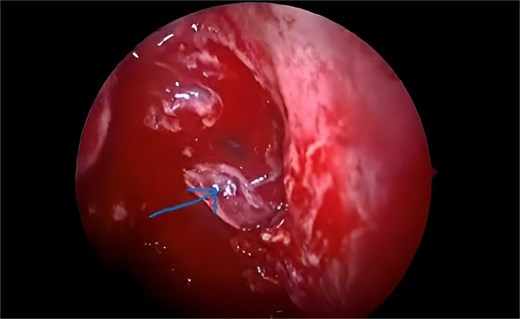
A 16-year-old boy was transferred to the hospital's otolaryngology (ENT) clinic complaining of a nasal voice and acute nasal dyspnea, and breathing difficulties, that had progressively grown over several months without the appearance of other symptoms. A clinical examination of the nose revealed a tumor that obstructed the right nostril and pushed the nasal septum to the far left. A computed tomography examination of the paranasal sinuses revealed that the tumor covered the whole right maxillary sinus and right nostril, preventing drainage from all sinuses on the same side and pushing the nasal septum to the opposite side. After the surgical intervention (Fig. 1) and endoscopic removal (Fig. 2), the nasal septum was restored to its original place. The removed sample was sent for analysis by a pathologist. The findings showed that the tumor was a maxillary sinus capillary hemangioma (Fig. 3). Due to the patient's low financial means, we chose not to do a magnetic resonance imaging (MRI). The patient was followed up for a month after surgery, and the Follow-up revealed that the patient's symptoms had reduced and that they were leading a normal life again.

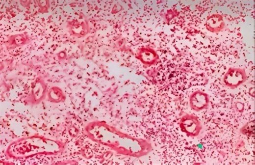
Capillary hemangioma of the maxillary sinus after excision.
A biopsy was performed on the mass, which is composed of two pieces of tan-colored tissue. The histopathological analysis reveals blood vessels of varying sizes, separated by fibrous septa. These vessels are lined with plump endothelial cells, and a few of them are filled with blood.
Maxillary sinus endoscopy showing hemangioma of the maxillary sinus.
Discussion
Hemangioma is a benign and rare vascular tumor that occurs in the facial sinuses. Accounting for only 10% of cervicofacial tumors [5]. To diagnose maxillary hemangiomas, we rely primarily on computed tomography (CT) and MRI [6]. In our situation, CT revealed a vascular lesion, but the diagnosis was not confirmed until postoperatively by histological examination of the mass. The patient was unable to perform an MRI due to financial constraints. The patient underwent successful endoscopic surgery with septal reconstruction. Endoscopic sinus surgery has proven to be an effective approach for managing maxillary sinus hemangiomas due to its low complication rates and rapid postoperative recovery [6, 7]. Alternative techniques, such as the pre-lacrimal approach, have also shown promising results [8]. A combined endoscopic procedure involving the Watsuji-Denker approach has been described in other reports [7–9]. Although preoperative embolization has been used in some cases to reduce the risk of intraoperative blood loss [10], in our instance, it was not necessary, as preoperative CT imaging suggested a non-hypervascular lesion, reducing the risk of severe intraoperative bleeding. This aligns with evidence indicating that well-planned endoscopic resection can achieve safe outcomes without the need for embolization [9–11]. Maxillary sinus hemangiomas are of two types: cavernous or capillary [12]. In our case, histological examination confirmed the diagnosis of hemangioma. Although it is a benign tumor, there is a high risk of recurrence or rare complications such as bleeding [13]. The patient's postoperative course was excellent, with no complications, recurrence, or localized swelling observed on follow-up imaging. Complete symptom resolution and return to normal activities were achieved, consistent with outcomes reported in similar cases [12, 13]. It is worth noting that this case represents the first reported case of maxillary sinus hemangioma in Syria, highlighting the rarity of such tumors in this region and supporting the feasibility of an exclusively endoscopic approach.
Conclusion
Rare vascular tumors and maxillary sinus hemangiomas demand precise diagnosis and treatment. While CT and MRI aid in preoperative assessment, histology remains essential for verification. The case demonstrates the benefits of endoscopic sinus surgery because it provides minimal invasion and fast recovery times with few adverse effects The careful planning of surgery allows most patients to avoid preoperative embolization procedures even though it might decrease bleeding in hypervascular tumors The case represents the first documented instance of this condition in Syria because such tumors are rare in this region and endoscopic treatment proved successful. Additional research must be conducted to create effective management approaches that will lead to better results for patients.
Acknowledgements
We want to thank the SMSR laboratory team for their efforts in bringing our team together.
Author contributions
Mais Alreem Basel Mohaisen: Corresponding author, writing the paper.
Conflict of interest statement
None declared.
Funding
None declared.
Provenance and peer review
This work was not commissioned and has been externally peer-reviewed.
Consent
Written informed consent was obtained from the patient's legal guardian.
Methods
The work has been reported per the SCARE criteria [14].
References
Sigdel B, Ghimire A, Parajuli R, et al.
