


Abstract
Lipoleiomyoma is a rare type of leiomyoma characterized by the presence of adipose tissue intermixed with smooth muscle fibers. Its clinical presentation is typically similar to those of typical leiomyomas. In this report, we describe the case of a 49-year-old asymptomatic woman whose histopathological examination confirmed the presence of uterine lipoleiomyoma.
Introduction
Lipoleiomyoma is an uncommon variant of leiomyoma characterized by the presence of adipose tissue interspersed within bundles of smooth muscle. Lipoleiomyoma has an estimated incidence between 0.03% and 0.2%. This tumor consists of benign smooth muscle intermingled with adipose tissue. Clinically, symptoms resemble those of conventional leiomyomas, ranging from asymptomatic cases to presentations with pelvic pain or vaginal bleeding. Although the uterine corpus is the most common site of occurrence, rare cases have been documented in the cervix, ovary, broad ligament, and retroperitoneum [1].
We report a case of a 49-year-old asymptomatic woman with an intrauterine device (IUD) who presented for a routine gynecological check-up and Pap smear. Imaging revealed a uterine mass initially suspected to be sarcoma; however, histopathological examination confirmed the diagnosis of uterine lipoleiomyoma.
Case presentation
A 49-year-old premenopausal woman with a history of C-section presented for a routine gynecological check-up and Pap smear at our hospital. She had been using Minera, a Levonorgestel IUD for contraception and reported no abnormal symptoms such as pelvic pain, abnormal bleeding, or weight loss. Physical examination was unremarkable.
Transvaginal ultrasound revealed a well-circumscribed, heterogeneous uterine mass with mixed echogenicity (Figs 1 and 2). Given the imaging characteristics, a malignant uterine neoplasm, particularly sarcoma, was initially suspected.
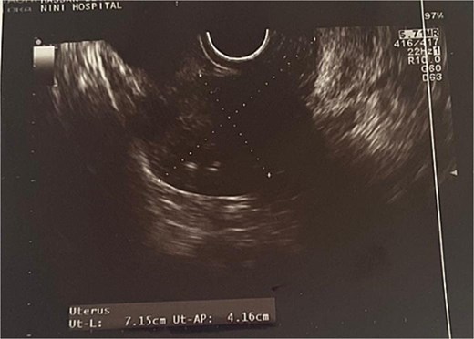
Transvaginal ultrasound showing the uterus.
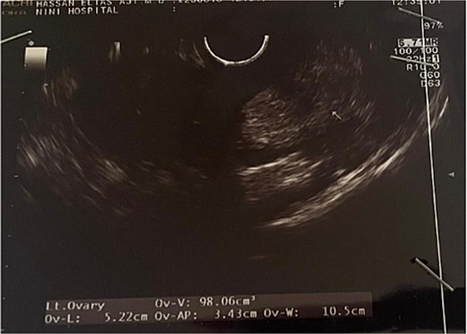
Transvaginal ultrasound showing the left ovary.
Magnetic resonance imaging (MRI) revealed an anterior uterine mass measuring 9.3 × 5.2 cm, with a posterior intramural component measuring 6.5 × 4.3 cm. The lesion was consistent with a subserosal-interstitial fibroid, corresponding to International Federation of Gynecology and Obstetrics classification Type 4–5: Type 4 being an intramural fibroid entirely confined within the myometrium, and Type 5 being a subserosal fibroid with more than 50% intramural extension. This mixed location accounts for the dual classification. The mass exhibited partial necrotic with a hemorrhagic area measuring 1.7 cm but demonstrated no radiological features suggestive of malignancy, such as irregular borders, invasive growth, or abnormal contrast enhancement.
Both ovaries were normal in size and morphology, with a few follicles up to 2 cm noted on the left. No pelvic lymphadenopathy or free fluid was identified. The primary finding was a benign uterine fibroid with areas of necrosis and hemorrhage. Due to these concerning radiologic features, the patient underwent a total abdominal hysterectomy and bilateral salpingo-oophorectomy. Histopathological analysis confirmed a myoma with lipomatous differentiation, without evidence of nuclear atypia or mitotic activity. The fallopian tubes were unremarkable, and a functional ovarian cyst was identified. Immunohistochemical staining was negative for malignancy-associated markers including Ki-67 (indicating a low proliferative index), p16, p53, S100, CD34, and HMB-45. Positive staining for desmin and smooth muscle actin confirmed smooth muscle origin. Collectively, these findings support a diagnosis of lipoleiomyoma and effectively rule out sarcoma, confirming the benign nature of the lesion.
Discussion
Lipoleiomyomas are rare tumors, often discovered incidentally during routine imaging. Their mixed composition can mimic malignant tumors such av liposarcoma or leiomyosarcoma, posing diagnostic challenges. Imaging techniques such as ultrasound and MRI play a crucial role in initial assessment, but a definitive diagnosis requires histopathological examination.
Malignant transformation is exceptionally rare, with only two reported cases of leiomyoliposarcoma [2].
While the majority of patients remain asymptomatic, some experience symptoms similar to those of typical leiomyomas, including vaginal bleeding, abdominal discomfort, pelvic and lower back pain, frequent urination, and constipation [3].
MRI with a fat-suppression sequence is especially useful for diagnosing uterine lipoleiomyoma due to its high sensitivity and specificity for detecting fat, as well as its ability to precisely locate the lesion through multiple sections. However, distinguishing uterine lipoleiomyoma from other uterine lipomatous tumors can sometimes be challenging. On macroscopic examination, the tumor typically presents as a well-defined, solid gray-tan mass with areas of yellow discoloration, indicating the presence of adipose tissue [4]. As mentioned previously, the standard treatment is hysterectomy.
Lipoleiomyomas are usually asymptomatic and clinically resemble leiomyomas, so they generally do not require specific treatment. The main surgical approach for these tumors is hysterectomy [5].
Lipoleiomyomas typically have a benign clinical course. However, there have been rare instances of lipoleiomyosarcomas developing from uterine lipoleiomyomas [6]. These can be differentiated from malignant forms by the relatively simple appearance of the nuclei and the occasional presence of mitosis in the smooth muscle component [7].
Conclusion
This case highlights the diagnostic complexity of lipoleiomyomas and the potential for being mistaken for malignant neoplasms. Accurate histopathological assessment is essential for distinguishing these benign lesions from sarcomas, ensuring appropriate management, and preventing overtreatment.
Lipoleiomyoma is an exceptionally rare tumor. Histologically, the presence of fat in the uterine tissue can be concerning, but it is limited to a few potential diagnoses, including benign conditions such as lipoma, lipoleiomyoma, teratoma, as well as malignant neoplasms. Liposarcoma may occur primarily or as part of sarcomatous transformation in mixed carcinoma. These lesions can typically be distinguished based on clinical and histological features [8].
Clinicians should consider lipoleiomyoma in the differential diagnosis of uterine masses, particularly in asymptomatic patients undergoing routine evaluations.
Conflict of interest statement
None declared.
Funding
None declared.
