-
PDF
- Split View
-
Views
-
Cite
Cite
Claudio Guerci, Andrea Kazemi Nava, Gloria Goi, Luca Ferrario, Francesco Cammarata, Giulia Lamperti, Piergiorgio Danelli, Post-traumatic diaphragmatic hernia: a rare case of intestinal obstruction, Journal of Surgical Case Reports, Volume 2025, Issue 3, March 2025, rjaf163, https://doi.org/10.1093/jscr/rjaf163
Close - Share Icon Share
Abstract
Post-traumatic diaphragmatic hernia is a rare and potentially life-threatening condition that can occur after blunt or penetrating trauma. Delayed presentations are uncommon, but can lead to serious complications, such as bowel obstruction. We report a case of a 31-year-old male patient who presented five years after a thoracic trauma with symptoms of intestinal obstruction and was diagnosed with a delayed post-traumatic diaphragmatic hernia. The diagnosis was made through contrast-enhanced computed tomography scan, and the patient underwent laparoscopic repair with mesh reinforcement. This case highlights the importance of considering diaphragmatic hernia in the differential diagnosis of patients with a history of trauma, even if the presentation is delayed. Prompt diagnosis and surgical intervention are crucial to prevent serious complications and improve patient outcomes. This study adds to the existing literature on traumatic diaphragmatic hernias, emphasizing the need for enhanced clinical awareness, interdisciplinary cooperation, and surgical repair.
Introduction
Post-traumatic diaphragmatic hernia is a defect in the diaphragm that allows the herniation of abdominal viscera into the thorax. It can result from blunt or penetrating trauma and pose a diagnostic and therapeutic challenge. Delayed presentations are uncommon but can lead to complications, like bowel obstruction [1]. Diagnosing diaphragmatic hernias after trauma might be delayed for different reasons: the absence of symptoms right away, other related injuries, or radiological findings misinterpretation [2, 3]. This can lead to diagnostic delay, with high morbidity and mortality rates. We report the case of a 31-year-old male patient with a five-year delayed presentation of post-traumatic diaphragmatic hernia.
Case report
This case report has been described in accordance with SCARE criteria [4].
A 31-year-old man was admitted to the emergency room for abdominal pain and vomiting, without stool and flatus. He had a history of thoracic trauma due to a stab wound, which caused a hemothorax and needed an urgent thoracotomy 5 years before (Fig. 1).
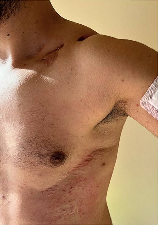
The ancient stab wound which caused a hemothorax and needed an urgent thoracotomy.
On physical examination, the abdomen was distended, along with a present bowel sound.
Biochemical parameters showed: CRP 118.7 mg/l leukocyte count 12 800 × 109/l; other values were normal.
The contrast-enhanced computed tomography (CT) scan revealed a left 2-cm-large diaphragmatic hernia at the level of the diaphragmatic dome, with herniation of the transverse colon and omentum (Fig. 2). There were no radiological signs of bowel obstruction.
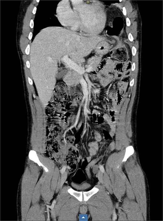
The patient underwent an initial non-operative management, with resolution of the sub-occlusion. Meanwhile, the patient was discharged, and surgery was programmed after 15 days.
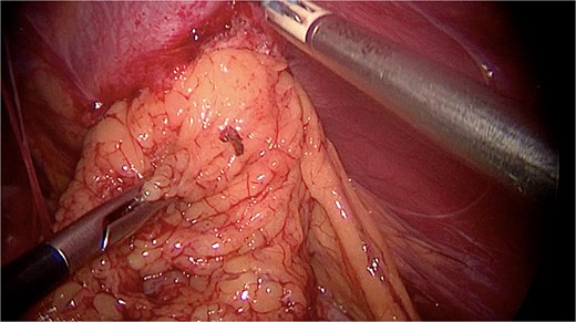
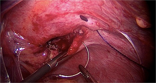
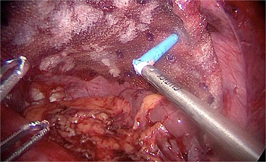
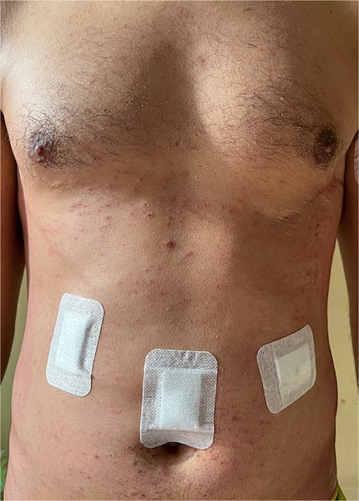
Under general anesthesia, a laparoscopy was made. Intraoperatively, a large diaphragmatic hernia was present with herniation of the omentum (Fig. 3). No transverse colon was found in the hernia, nor were signs of bowel occlusion documented. The content of the hernia was reduced into the peritoneal cavity after performing adhesiolysis and checking organ vitality. The hernia cavity was totally extra-pleural. The parietal pleura was localized cranially, and no expansion was documented even after pulmonary recruitment. The defect in the diaphragm was sutured with PDS 1 go-and-back suture (Stratafix) (Fig. 4) and then covered by a biosynthetic prosthesis (Phasix Mesh), fixed by AbsorbaTack and cyanoacrylate glue (Fig. 5). An intra-abdominal drain was placed. Following the surgery, the patient recovered well and was discharged on the fourth postoperative day (Fig. 6). No postoperative complications occurred. The postoperative imaging control was normal. At the six-month follow-up, the patient showed no signs of recurrence.
Discussion
The displacement of intra-abdominal organs into the chest through a diaphragmatic orifice caused by trauma is known as a post-traumatic diaphragmatic hernia. While its early manifestation after trauma is widely documented, cases of delayed presentation with consequences such as intestinal obstruction are uncommon and challenging. In as many as 60% of instances, the first imaging fails to detect the diagnosis of diaphragm rupture with forceful trauma [5]. Traumatic diaphragm rupture may be undiagnosed in an emergency because of other serious injuries [6]. Diaphragmatic hernia results from failing to notice the diaphragm rupture, and it might develop months or even years later, rarely with symptoms of intestinal obstruction [7]. Traumatic diaphragmatic hernia is primarily caused by blunt trauma, with the left hemidiaphragm being the commonly damaged [8].
In our case, the patient's history of thoracotomy-related trauma five years before presentation raised concerns about the delayed onset of a diaphragmatic hernia [9].
The best imaging method for identifying diaphragmatic injuries is computed tomography, which also provides useful information for surgery planning [10].
Traumatic diaphragmatic hernias are divided into three categories: the diagnosis of type 1 hernia is made right after the event. Type 2 is diagnosed during the recovery period. A type 3 is diagnosed when a patient exhibits signs of herniated organs [11]. Our case is classified as Type 3.
The gold standard procedure for repairing diaphragmatic and visceral injuries in emergency is exploratory laparotomy. The decision about the best surgical approach depends on the chronicity of the condition, the surgeon’s preferences and skills, and the local resources [12]. Since there was a possibility of adhesions in the abdominal portion of the herniated colon, a laparoscopic abdominal approach was recommended in our case. Anyway, laparoscopic approach has become the most used approach to manage complicated diaphragmatic hernias [13]. Robotic surgery of complicated DH repair has been reported only in a few cases [14].
Reducing herniated organs and fixing the diaphragmatic defect are two key steps. Restoring normal anatomy and preventing further complications were the goals of this surgical strategy. This is why we opted for mesh positioning.
The reconstruction can be performed with synthetic meshes, which are well tolerated, bio-prosthetic materials or entirely artificial mesh, either absorbable or non-absorbable [12]. Biological meshes have a lower rate of hernia recurrence, higher resistance to infections and lower risk of displacement [15]. In our case, we decided to reinforce the diaphragmatic defect with a biosynthetic mesh (Phasix) since it is demonstrated to be safe and effective [16]. It leads to lower recurrence rates with a quality-of-life improvement in diaphragmatic hernia repair [17].
This case report focuses on some important aspects:
First, intestinal obstruction-complicated delayed presentation is a rare and difficult situation [18]. The need of taking diaphragmatic hernia into account in the differential diagnosis is highlighted by the existence of a previous history of trauma.
Second, the radiological results were crucial in establishing the diagnosis and directing the surgical decision-making. This case's rarity emphasizes the value of clinical awareness and interdisciplinary cooperation [19].
Third, this case report adds to the body of knowledge already available on traumatic diaphragmatic hernias, namely the delayed manifestation of intestinal obstruction and the safety and effectiveness of mesh reinforcement.
In conclusion, delayed traumatic diaphragmatic hernias, which can be misdiagnosed, are uncommon but potentially fatal illnesses that need to be identified and treated. This case highlights how crucial it is to take diaphragmatic hernias into account in patients who have had trauma, even if their presentation is delayed. Diagnosing diaphragmatic injuries requires CT scans, and the mainstay of treatment is still surgery, which includes repairing the diaphragmatic defect and reducing herniated organs.
Conflict of interest statement
All authors declare that they have no conflict of interest. Informed consent was obtained from the patients being included in the study.
Funding
None declared.
Ethic statement
Ethical review and approval were not required for the study on human participants in accordance with the local legislation and institutional requirements. The patients/participants provided their written informed consent to participate in this study. Written informed consent was obtained from the individual(s) for the publication of any potentially identifiable images or data included in this article.
References
Kumar S, Kumar S, Bhaduri S, et al. An undiagnosed left sided traumatic diaphragmatic hernia presenting as small intestinal strangulation: a case report. Int J Surg Case Rep 2013;4:446–8.


