


Abstract
High pressure injection injuries to upper extremities are largely rare occurrences. The direct and indirect effects of the injected substance can cause debilitating consequences for those affected and can lead to digit amputation. This case series includes three patients with high pressure injection injuries admitted to a single tertiary centre over one year. One patient was discharged from both plastic surgery and hand therapy 6 months post injury. He reports good function but has residual numbness and cold intolerance. The second individual is still linked to surgical outpatients and has ongoing hand therapy requirements, stiffness, and numbness 12 months post injury. The final patient represented three months post initial debridement with an infected finger that required amputation. Based on these findings, it is imperative that prompt surgical debridement occurs in conjunction with early hand therapy intervention to achieve optimal outcomes for patients.
Introduction
Hand injuries are common presentations to emergency departments globally and those requiring admission include infections, fractures, lacerated tendons and nerves as well as more complex wounds [1].
However, high pressure injection injuries of digits are fortunately uncommon. Such occurrences are considered to be surgical emergencies. There is a general consensus that early surgical exploration is necessary to decompress the digit [2, 3] but there are no standardized guidelines. The decision to aggressively debride the injected substance to reduce the cascade of inflammation and tissue destruction is made on a case by case basis. The first report of such an injury was documented in Germany in 1925 [4]. Subsequently, Rees in 1937 reported a case whereby a mechanic endured severe infection following a high pressure injection with diesel that resulted in amputation [5].
Despite the passage of nearly 100 years since the aforementioned accidents, the significant developments in surgery and advances in healthcare, amputation and severe dysfunction are not uncommon outcomes post such injuries even today.
Presentation
The most common presentation of a high-pressure injection injury is a puncture wound on the nondominant hand of a male manual worker [6]. The most frequently cited causative agents include oil, paint, chemicals, and air [7]. The ensuing damage post injury is dependent upon a number of factors including the nature and volume of the substance, the force with which it is introduced, the timing and extent of surgical debridement and the presence of subsequent infection [8].
Pathophysiology
The tissue destruction associated with high pressure hand injuries can be considered as having three phases. The first phase relates to the direct mechanical impact of injection with resultant swelling, consequent pressure-induced neurovascular compromise and potential compartment syndrome. The second stage is driven by the cytotoxic or vesicant effects and the subsequent inflammatory response the agent has on tissues. The third phase relates to potential secondary infection caused by microbial contamination at the time of injury [9]. There are varied reports on the risk of infection during the initial injury and post debridement but there is a general consensus that patients with high pressure injuries should be commenced on broad spectrum antibiotics [10].
Case series
This case series includes all high pressure injection injuries to digits admitted to a tertiary centre over a one year period. All three patients were male manual workers with high pressure injuries from paint. Two of the patients were operated on within 10 hours of presentation to hospital but one individual did wait over 24 hours. This was unfortunately due to delayed theatre access. This individual had a protracted course of recovery with the most re admissions. One man had his digit amputated. Table one outlines the presentation and outcomes of all three patients in this study (Table 1).
Patient demographics and parameters associated with the injuries
| Patient one | Patient two | Patient three | |
|---|---|---|---|
| Hand dominance | Left hand | Right hand | Right hand |
| Washouts required during initial stay in hospital | Washout Day 2, 3, 7, 9, 10 (BTM applied during initial admission due to exposed tendon) | 2 within 72 hours | 2 within 72 hours |
| Age | 27 | 55 | 31 |
| Digit affected | Left middle | Right middle | Left index |
| Entry point | Middle phalanx volar surface | Pulp at distal tip | PIPJ at the ulnar border |
| Clinical presentation | Digit not swollen Painful FDS/FDP intact – reduced sensation in both nerve bundles | Swollen digit Painful FDS/FDP intact – reduced sensation in both nerve bundles | Swollen digit Painful FDS/FDP intact – reduced sensation in both nerve bundles |
| X-ray at presentation | Soft tissue swelling with increased density | Soft tissue swelling with increased density | Soft tissue swelling with increased density |
| Time to theatre from arrival at hospital | Over 24 hours | Within 4 hours | Within 10 hours |
| Treatment | Decompression and debridement | Aggressive debridement of entire digit | Decompression and debridement |
| IV antibiotics | Clindamycin and co-amoxiclav | Co-amoxiclav | Co-amoxiclav |
| Length of stay post injury (days) | 11 | 4 | 11 |
| Readmissions | Yes 2 (1 acute, 1 elective) Readmitted 44 days post injury with infection. He was treated with antibiotics and BTM reapplied Elective admission 98 days post injury for BTM delamination and skin graft | No | Yes (1 elective) Readmitted 65 days post injury for elective terminalization of digit at DIPJ following a visit to outpatient 1 week previous with stiff hand and pus exudate from original wound. |
| Outcomes | Stiff finger, residual pain. | Numbness and returned to work within 2 weeks | Numbness post initial debridement Amputation post readmission at level of DIPJ |
BTM, biodegradable temporising matrix; DIPJ, distal interphalangeal joint; FDP, flexor digitorum profundus; FDS, flexor digitorum superficialis; PIPJ, proximal interphangeal joint.
Patient one
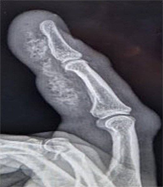
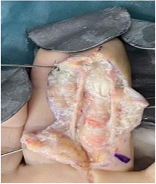
Initial presentation (Fig. 1) with accompanying X-ray (Fig. 2) and intra op image during first debridement (Fig. 3).
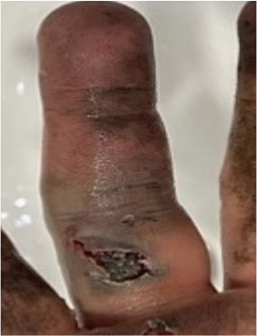
Image of patient one, showing his digit at presentation displaying large puncture wound on volar surface.
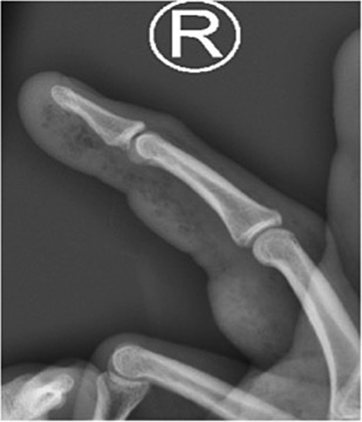
X-ray image of patient one showing injected material into volar surface of his digit.
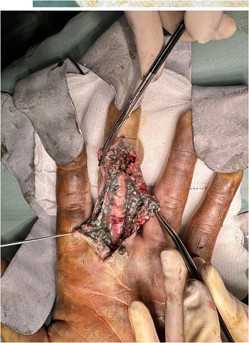
Intra op image of patient one’s original debridement.
Patient two
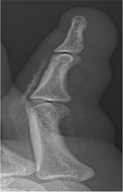
Initial presentation (Fig. 4) with accompanying X-ray (Fig. 5) and intra op image during first debridement (Fig. 6).
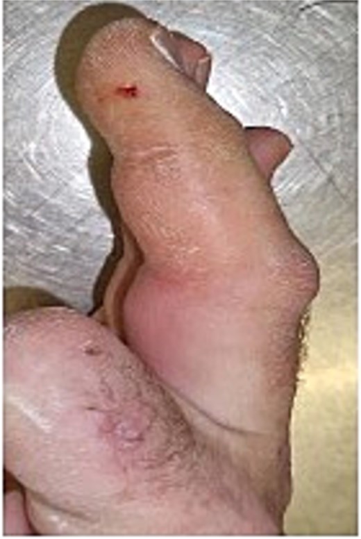
Image of patient two, showing his digit at presentation displaying very small puncture wound on lateral volar surface of his digit.
X-ray image of patient two showing injected material into volar surface of his digit.
Intra op image of patient two’s original debridement.
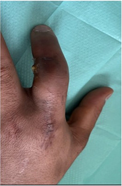
Patient three
Initial presentation (Fig. 7) with accompanying X-ray (Fig. 8). Final image shows patients digit when he represented 2 months after original insult with a swollen finger and pus exudate (Fig. 9).
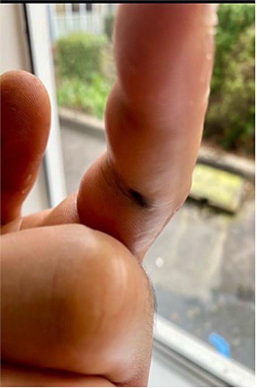
Image of patient three, showing his digit at presentation displaying small puncture wound on volar surface.
X-ray image of patient three showing injected material into surface of his digit, more obvious in the proximal portion.
Patients three’s digit when he represented 2 months after original insult with a swollen finger in a flexed position and pus exudate.
Discussion
This case series, adds to the current body of evidence that advocates for early decompression and debridement of these high pressure injuries. Initial treatment should also include tetanus prophylaxis (if indicated) with the addition of broad-spectrum antibiotics pre theatre. For all three patients, our specialized hand occupational therapists were involved in rehabiliation immediately in the post operative period and this is an element of care that is imperative to optimize outcomes for patients.
Despite empirical antibiotic treatment for all patients, two patients were re admitted with infections and one man required amputation almost 10 weeks post injury. As mentioned previously, terminalization of a digit is not uncommon post such injuries. We would recommend that routine wound swabs be taken during all surgical interventions or at any juncture that infection is suspected.
One review over a 28 year period ending in 2000, catalogued 43 high pressure injuries and found 12 patients (28%) required amputation and only 9 reported no functional deficit in the aftermath of the accident [11]. Another review conducted by Feldman et al. included eight people in one centre and reported that all patients treated, reported from some level of neuropathic pain and/or cold intolerance post inoculation with a substance under high pressure conditions [12]. This is reflected in our data set whereby all patients endured residual pain or numbness post injury.
Comparing the three individuals described in this review, the patient that had the best outcome was the one that was taken to theatre within 4 hrs of presentation at the hospital and who had immediate debridement of all visible material. A retrospective review of 11 high pressure injuries in a UK single centre emphasized the high morbidity associated with paint gun injuries [13]. We would therefore advocate for this treatment algorithm of early decompression and debridement for high pressure injuries involving paint. Other authors have advocated for consideration of early amputation in the presence of a digit that is cool or poorly perfused at presentation [14].
The patient that ultimately had a delayed terminalization was not compliant with aftercare instructions or outpatient appointments. This emphasizes the fact that intense support of these individuals is needed not only in the acute post op period but also in the weeks and months post injury. The pivotal role of hand therapist cannot be under estimated. The literature recognizes the need to maintain good range of motion of the affected hand from the outset [6] and this can certainly be achieved with early referral to therapist colleagues.
Undoubtedly, patients are autonomous and despite the best efforts of teams some don’t adhere to post op advice regarding surgical and hand therapy follow up for a variety of reasons. All clinicians and therapist can do is emphasize to all those affected by high pressure injuries the seriousness of the insult and the critical need to engage with services post discharge.
Conclusion
In the acute stage of these injuries early aggressive debridement is the cornerstone of treatment but close monitoring post op to evaluate the need for further washouts in conjunction with hand therapy is as beneficial to the patient in terms of reducing their risk of long term negative sequela.
Conflict of interest statement
None declared.
Funding
None declared.
