


Abstract
The mandible is one of the most frequently fractured facial bone, most commonly by automobile accidents followed by fall from heights. However, segmental avulsion of mandible, that too in pediatric population is rarely reported. We report a case of 10-year-old male child sustaining facial injury following fall from a tree, resulting in complete segmental avulsion of the mandible highlighting the importance of early plastic surgery referral to optimize treatment outcomes. After clinical and radiological evaluation, the patient underwent free fibula osseocutaneous flap reconstruction. Given the greater healing potential of facial bones in younger patients, re-fixation of the avulsed segment would have been considered if presenting well within time.
Introduction
Less than 15% of all facial fractures occur in children and 1% of them occur in children under 5 years of age [1]. Among various etiologies, fall from height is very common in this age group [2]. The reported incidence of mandibular fractures is ~20%–50% of all childhood facial fractures [3]. The most frequent site of pediatric mandibular fractures is the condylar region, followed by the symphysis/para symphysis, angle, and body, respectively [4]. The mandible being a U-shaped bone, has horizontal and vertical segments along with strong muscular attachments that are responsible for various movements and determine the displacement of segments after trauma. However, it is very unlikely that there is total avulsion of the mandibular segment, that too in the pediatric age group [5]. The mandible significantly defines the lower third of the face and is important for mastication hence careful repair and restoration of function is an important task while managing them. Considering the dynamic nature of pediatric craniofacial growth, management of facial fractures in this age group requires careful consideration to avoid long-term deformities and functional impairments [6]. This article highlights on gap in the current understanding regarding effective management of pediatric mandibular avulsion and emphasizes on timely referral to plastic surgery to optimize treatment outcomes.
Case presentation
A 10-year-old male sustained facial injury and bleeding from mouth following accidental fall facing the ground from a tree approximately 10 feet in height (Fig. 1). No other body injury was reported. The patient was rushed to a nearby hospital within an hour of trauma, where primary treatment was done. On referral to a higher center for further management, loss of right-sided avulsed mandibular segment was seen and on inquiry, the lost segment was retrieved from the incident site almost after 12 hours of trauma (Fig. 2). However, the segment was discarded because of non-viability, facial laceration was closed primarily and Ryle’s tube (RT) was inserted. The patient was then referred to us for mandibular reconstruction.
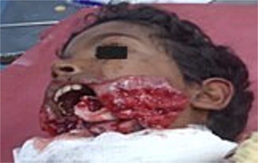
Patient at time of initial presentation with open wound.
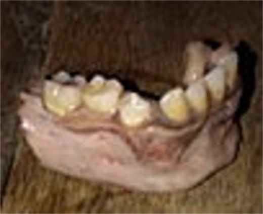
Avulsed segment of mandible.
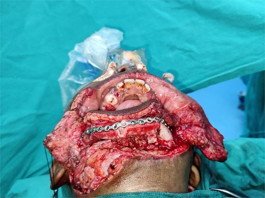
The patient presented to our tertiary center after 2 weeks of injury with complaints of inability to close mouth completely, and continuous drooling of saliva along with difficulty in chewing. On examination facial asymmetry was noted in the lower third (Fig. 3), normal occlusion was absent with loss of all four lower incisors, right canine, and two premolars along with obliteration of gingivobuccal sulcus on the right side (Fig. 4). However, cant of oral commissure was at same level, lip seal was adequate and mouth opening was normal. Facial nerve and bilateral condylar movements were also normal. There was a healed scar along the mandible extending from the left premolar region to 3 cm anterior to angle of mandible on right side (Fig. 5). On further investigation with non-contrast computed tomography (NCCT) face with 3D reconstruction, segmental loss of the body and symphysis on right side of mandible was seen (Fig. 6).
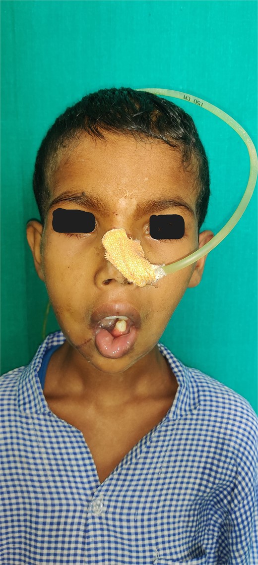
Clinical image of patient showing inability to close mouth completely.
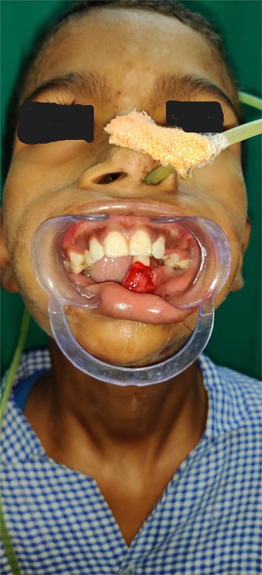
Intra-oral clinical image showing lost mandibular segment.
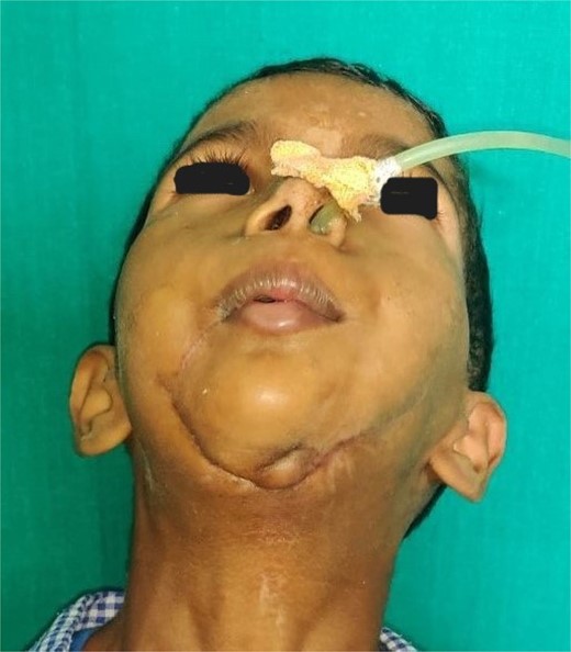
Clinical image showing healed scar over chin indicating site of primary closure.
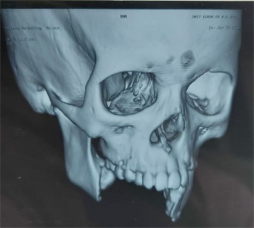
NCCT face with 3D reconstruction showing loss of symphysis and right-side body of the mandible.
In view of functional restoration and dental rehabilitation, the lost segment was planned to be reconstructed with a free fibular osseocutaneous flap, and consent for the same was taken from family members after explaining to them the procedure and its outcomes. Preoperative perforator marking for skin paddle was done through a handheld Doppler (Fig. 7). Intraoperatively the skin flap at previously sutured scar was raised and the facial artery and external jugular vein on right side were dissected and prepared for microvascular anastomosis. The actual bony defect was measured to be 6 cm and a free fibula flap with 7*5 cm of skin paddle was harvested from the left leg (Fig. 8). Leaving behind 6 cm each of proximal and distal fibular segments for knee and ankle stability, respectively, remaining length of fibula is usually harvested in view of gain in length of pedicle. We did not use any three-dimensional printing technology for defect assessment and planning due to financial constraints. A single closed wedge osteotomy (2 cm) was made to create 2 cm of body and 4 cm of symphysis. Bony fixation was done with a large 16-hole titanium reconstruction plate (Fig. 9). Intraoperatively mouth opening of 4 cm was achieved. Donor site was closed primarily and a below-knee plaster of Paris (POP) slab was applied.
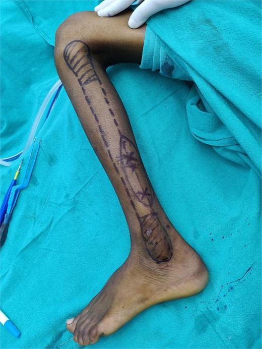
Preoperative marking of peroneal artery perforators using hand-held Doppler along with skin paddle.
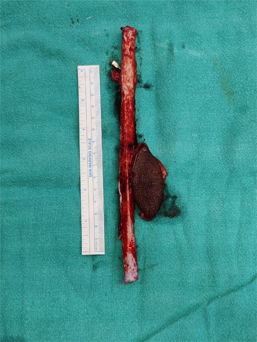
Harvested fibula along with skin paddle.
Fixation of free fibula flap with a 16-hole titanium plate.
The patient was monitored in intensive care unit (ICU) for a total of 3 days with RT in situ. RT feed was initiated on second day after surgery, and controlled ambulation was gradually allowed on fifth postoperative day. The patient was able to open and close his mouth completely with an inter-incisor distance of 2.5 cm (Fig. 10). The facial sutures were removed on day 7 and the patient was discharged. The donor site sutures and POP slab were removed on day 14 of surgery following which the patient was allowed for weight-bearing ambulation and oral feeds. In further follow-up visits, scar care was done and no donor site complications were recorded. A secondary dental rehabilitation was planned using Osseo-integrated titanium implants.
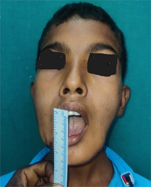
Postoperative image showing mouth opening.
Discussion
Complete avulsion of the mandibular segment as in present case is rarely reported incident [7]. There are several studies emphasizing refixation of the avulsed bony segment, especially in young patients whenever possible due to higher healing potential of facial bones. Younas et al. have reported healing of a large avulsed mandibular segment following re-fixation [5]. The use of non-vascularized bone graft for a length of up to 5 cm is also well-established [8]. In this case, as the defect was 6 cm and the avulsed bony segment was not available for re-fixation, reconstruction with a free fibula flap was planned, and adequate mouth opening along with retur n of facial contour was achieved. A study on mandibular reconstruction in pediatric patients with free fibula flap has reported early return of function and psychosocial stability without any associated morbidities [9].
Conclusion
Reconstruction of lost mandible is not only desirable but indispensable, that too in a child. Re-fixation is one good option provided the patient presents within time, however, a free fibula osseocutaneous flap remains the gold standard of treatment. In both scenarios, the role of plastic surgeons in definitive management of mandibular avulsion is indispensable for early restoration of function and esthetics.
Guarantor
Dr. Anupama Kumari, M.Ch, Department of Burns and Plastic Surgery, All India Institute of Medical Sciences, PATNA, India, Mobile: 8795381249, Email: anu786.doc@gmail.com
