


Abstract
Amyand’s hernia, defined as the presence of the vermiform appendix within an inguinal hernia sac, is a rare condition, particularly in neonates. This report describes a 1-month-old preterm male presenting with scrotal swelling, vomiting, watery stools, and excessive crying. Examination revealed abdominal distension and an irreducible, tender right groin swelling, initially diagnosed as an obstructed inguinal hernia. Intraoperative findings confirmed a perforated appendix within the hernia sac, necessitating appendectomy and herniotomy. The patient recovered uneventfully, resuming feeding within 24 h and being discharged 48 h postoperatively with normal bowel function. This case underscores the diagnostic challenges and successful surgical management of perforated Amyand’s hernia in a neonate, highlighting the importance of prompt intervention to prevent complications.
Introduction
Amyand’s hernia, first described by Claudius Amyand in 1735, occurs when the vermiform appendix is found within an inguinal hernia sac. Its incidence ranges from 0.19% to 1.7%, with acute appendicitis complicating ~0.07% to 0.13%. The condition is three times more prevalent in children than adults, often linked to a patent processus vaginalis [1]. Perforation of the appendix within the sac, classified as Type 3 per Losanoff and Basson’s system, is exceedingly rare and carries a mortality risk of 15% to 30% [1, 2]. This classification categorizes Amyand’s hernia into four types: Type 1 (normal appendix), Type 2 (appendicitis confined to the sac), Type 3 (appendicitis with peritonitis), and Type 4 (appendicitis with additional abdominal pathology) [2].
In neonates, Amyand’s hernia often presents as an incarcerated or strangulated hernia, mimicking conditions like testicular torsion or orchitis, which complicates preoperative diagnosis [3]. Imaging such as ultrasound may assist but is often inconclusive, leading to intraoperative confirmation [4]. This report details a rare case of perforated Amyand’s hernia in a preterm neonate, emphasizing clinical presentation, surgical management, and outcomes.
Case report
A 1-month-old male, born preterm at 32 weeks gestation, presented with a 1-day history of right scrotal swelling, vomiting, watery stools, and excessive crying. On examination, the infant was in fair general condition with abdominal distension, hyperresonant percussion, high-pitched bowel sounds, and a firm, irreducible, and tender right groin swelling. A diagnosis of obstructed right inguinal hernia was established. Informed consent was obtained for emergency herniotomy.
Intraoperatively, through a right inguinal incision, the hernia sac contents could not be reduced (Fig. 1). Upon opening the sac, a perforated appendix was identified (Fig. 2). An appendectomy was performed, the cecum was reduced into the abdomen, high ligation of the sac was performed, and the abdominal wall closed in layers. The patient resumed feeding 24 h postoperatively and was discharged within 48 h with normal bowel function.
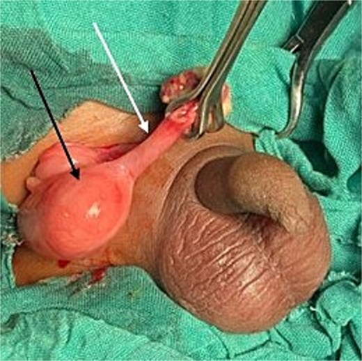
Intraoperative view of Amyand’s hernia. Black arrow: cecum; white arrow: inflamed appendix.
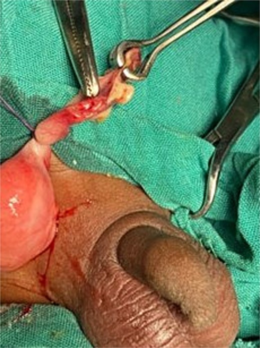
Intraoperative view of perforated appendix.
Discussion
Amyand’s hernia, characterized by the incarceration of the vermiform appendix within an inguinal hernia sac, represents a rare surgical entity, particularly in the neonatal population [1]. The incidence of appendicitis complicating Amyand’s hernia is rare, with perforation occurring in an estimated 0.1% of cases [1, 2]. In neonates, the condition is exceedingly uncommon, with most reported instances involving premature babies and linked to a patent processus vaginalis [5]. The pathophysiology remains incompletely understood but is thought to involve mechanical compression of the appendix within the hernia sac, leading to vascular compromise, ischemia, inflammation, and eventual perforation [6]. Additional factors in neonates, such as increased intra-abdominal pressure from crying or feeding issues, may exacerbate this process [7].
Diagnostic challenges are profound in neonatal Amyand’s hernia, particularly when perforated, as the presentation often mimics common conditions such as strangulated inguinal hernia, testicular torsion, epididymo-orchitis, or acute scrotum [8]. In this case, the presenting symptoms led to a preoperative diagnosis of obstructed inguinal hernia. Similar nonspecific symptoms have been reported in other cases, where fever, irritability, and leukocytosis may be present but are not pathognomonic [6]. Sandhu et al. report a case of a 25-day-old neonate with inguinoscrotal swelling, bilious vomiting, and dehydration subsequently diagnosed with a perforated Amyand’s hernia [7]. Preoperative imaging, such as ultrasound, can occasionally aid in identifying the appendix within the sac or detecting fluid collections suggestive of perforation, but it is inconclusive and may be omitted in emergent settings [9]. Computed tomography is rarely used in neonates due to radiation concerns, though it has been advocated in ambiguous adult cases [3]. The reliance on clinical suspicion underscores the need for a high index of awareness among clinicians, as delayed diagnosis can result in complications like scrotal abscess or peritonitis [10].
According to Losanoff and Basson’s classification, this case qualifies as a Type 3 Amyand’s hernia, involving acute appendicitis with peritonitis or perforation extending beyond the sac [2]. Type 1 hernias require hernia repair alone, while Types 2–4 require appendectomy due to inflammation or perforation [1]. In neonates, surgical intervention is invariably through an inguinal approach, with appendectomy, reduction of viable bowel, and herniotomy via high ligation of the sac. Mesh reinforcement is contraindicated in contaminated fields [11]. Debate exists regarding prophylactic appendectomy for normal appendices in Amyand’s hernia, with some arguing against it in children to preserve the appendix for potential future reconstructive uses, such as urinary diversion [12]. However, with perforation, appendectomy is mandatory, often accompanied by broad-spectrum antibiotics, thorough irrigation, and drainage to mitigate sepsis risks [7]. Laparoscopy has been described in older children and adults [3], but its application is limited in low-resource settings.
Our case is similar to other reports in the literature. Abdullateef et al. report successful treatment of a perforated Amyand’s hernia in a 26-day-old neonate [13]. Similarly, a premature infant with perforated Amyand’s hernia developed post-operative bowel necrosis and mesenteric thrombosis, highlighting potential complications in extremely preterm babies [14]. Another preterm case complicated by scrotal abscess emphasized containment of infection within the sac, which may explain better outcomes compared to intra-abdominal perforations [10]. In a series of infantile cases, timely surgical exploration led to complication-free recoveries, reinforcing the value of prompt intervention [9]. Amyand’s hernias are predominantly right-sided, though left-sided variants may indicate underlying anomalies like situs inversus [7]. Neonatal appendicitis within Amyand’s hernia show a male preponderance (9:1 ratio) and higher perforation rates due to immature immune responses and thin appendiceal walls [13].
Outcomes are generally favorable with early surgery, as evidenced by this patient’s prompt resumption of feeding and discharge within 48 h. However, delays can lead to severe sequelae, including sepsis or abscess formation, with mortality up to 30% in perforated neonatal cases [6]. Long-term follow-up is essential to monitor for hernia recurrence, though most reports indicate symptom-free periods post-discharge [8]. Future directions may include enhanced preoperative diagnostics via point-of-care ultrasound or assisted imaging analysis to improve detection rates, potentially reducing operative surprises [13]. This case contributes to the sparse literature, advocating for routine sac exploration during neonatal hernia repairs and heightened vigilance in preterm infants with irreducible groin masses.
Conclusion
This case highlights the successful management of a perforated Amyand’s hernia in a preterm neonate through emergency appendectomy and herniotomy. The nonspecific presentation underscores the need for a high index of suspicion in infants with irreducible groin swellings. Early surgical exploration, guided by clinical findings and supported by imaging when feasible, is essential to mitigate life-threatening complications. This report adds to the limited literature on neonatal Amyand’s hernia, emphasizing the importance of prompt diagnosis and tailored surgical intervention.
Conflict of interest statement
None declared.
Funding
None declared.
