


Abstract
Ventriculoperitoneal shunting, though effective for hydrocephalus, can rarely cause distal catheter migration. Scrotal migration via a patent processus vaginalis is well known in children but remains uncommon and underreported. We report a 6-month-old male who underwent right-sided lumboperitoneal-ventriculoperitonea (VP) shunt placement after neonatal meningitis at 10 days old. At 2 months, he presented with a left inguinal swelling. Examination showed a firm, tender, non-transilluminant mass suggestive of hernia, with the shunt functioning and the child stable. Surgery confirmed VP shunt catheter herniation into the scrotum. The catheter was reduced into the peritoneal cavity using a non-touch technique, and the hernia was repaired without complications. Recovery was uneventful, and he was discharged the next day. This case underscores the need for heightened clinical vigilance in infants with VP shunts who present with inguinal swelling. Multidisciplinary surgical intervention ensures favourable outcomes.
Introduction
Ventriculoperitoneal (VP) shunting remains the gold standard for managing hydrocephalus, a condition characterized by the pathological accumulation of cerebrospinal fluid (CSF) within the brain’s ventricles. The procedure involves the diversion of CSF from the ventricles to the peritoneal cavity, thereby alleviating intracranial pressure. Despite its widespread use and effectiveness, VP shunting is not without complications. Reported issues include infection, obstruction, over-drainage, and mechanical problems such as disconnection, fracture, or migration of the catheter components [1].
One of the rarer mechanical complications is the distal migration of the peritoneal catheter into the scrotum—a phenomenon facilitated by a patent processus vaginalis (PPV). This congenital anomaly is more prevalent in infants and preterm neonates, with persistence rates of up to 90% in preterm males and ~20%–30% in full-term males [2, 3]. The PPV, a peritoneal diverticulum accompanying the testis during its descent, typically obliterates after birth. Failure of closure creates a conduit through which intra-abdominal structures, including VP shunt catheters, may herniate.
Scrotal migration, though rare and often underreported, can cause shunt malfunction, testicular damage, and infection. Its symptoms may resemble inguinal hernia or hydrocele, leading to delayed diagnosis [4, 5]. This report adds to the limited body of literature and highlights the importance of early recognition and interdisciplinary management in such cases.
Case report
A 6-month-old male infant was brought to the paediatric clinic by his parents due to a swelling in the left inguinal region. The infant had a history of neonatal meningitis and underwent right-sided lumboperitoneal to ventriculoperitoneal (LP-VP) shunt placement. Since the shunt procedure, the child had shown steady neurological development and had not exhibited signs of increased intracranial pressure.
At presentation, the infant was active, alert, afebrile, and feeding well. There were no symptoms of vomiting, irritability, or refusal to feed. The VP shunt appeared to be functioning clinically, with no signs of malfunction such as lethargy, bulging fontanelles, or seizures.
On physical examination, a firm, smooth, non-fluctuant, non-transilluminant swelling measuring ~2 × 3 cm was noted in the left inguinal region. The mass was tender on palpation but did not exhibit signs of inflammation. No abdominal masses were palpable. Based on clinical findings, an inguinal hernia was suspected (Fig. 1).
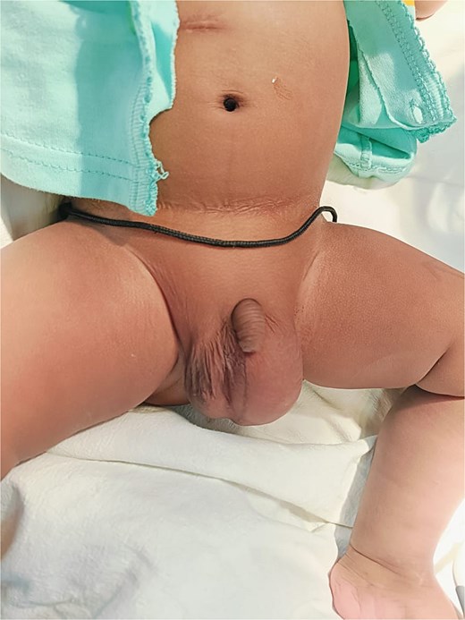
Clinical findings.
Basic laboratory investigations including a complete blood count and inflammatory markers were within normal limits. Due to the classic presentation and the urgency of the case, imaging such as abdominal ultrasonography or X-ray was deferred, and surgical repair was planned.
The patient was taken to the operating room for elective left inguinal hernia repair under general anesthesia. After positioning the infant in the supine position, a lower abdominal transverse incision was made on the left side.
The external oblique aponeurosis was incised to access the inguinal canal. The vas deferens and testicular vessels were identified and preserved with care. Upon examination of the hernial sac, the distal end of the VP shunt catheter was found entering the scrotum through the patent processus vaginalis. The hernia sac was carefully opened, and the shunt catheter was visualized within the scrotal cavity. Using a non-touch technique, the catheter was gently reduced back into the peritoneal cavity (Fig. 2). The hernia sac was ligated high and closed using absorbable sutures (Vicryl). The surgical wound was closed in layers, with the skin approximated using Monocryl. The procedure was completed successfully without any intraoperative complications. The infant recovered well post operatively and was discharged on the next day with a scheduled follow-up.
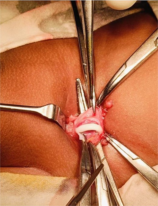
Intraoperative image showing VP shunt entering the scrotum.
Discussion
Scrotal migration of the VP shunt is an infrequent but clinically significant complication, especially in the pediatric population. The pathophysiology is primarily anatomical and mechanical—rooted in the presence of a PPV. The ongoing patency allows the distal end of the VP shunt to traverse from the peritoneal cavity into the inguinal canal and scrotum, a pathway that becomes more accessible with increased intra-abdominal pressure from CSF outflow [1–3].
Our case illustrates a left-sided scrotal migration in a patient with a right-sided VP shunt, emphasizing that migration can occur contralaterally. This supports the notion of bilateral PPV or dynamic reopening under persistent CSF pressure [4]. This case also underlines the importance of considering scrotal swelling as a possible presentation of shunt migration rather than defaulting to common pediatric diagnoses like hernia or hydrocele.
A review of published literature reveals only sporadic reports and small case series addressing this complication. Hauser et al. compiled 48 cases of scrotal VP shunt migration, suggesting that while rare, the phenomenon warrants systematic attention and possibly preventive strategies such as preoperative hernia screening and prophylactic herniotomy in high-risk infants [5].
Surgical management generally involves reduction of the catheter and high ligation of the hernia sac. In our case, we adopted a non-touch technique to prevent iatrogenic contamination, followed by a standard hernia repair. The absence of shunt malfunction or infection allowed us to avoid more extensive interventions such as shunt revision or externalization [2, 4].
Failure to address this condition early may result in serious consequences including catheter infection, CSF leak, testicular ischemia, or mechanical dysfunction of the shunt system. As seen in our patient, timely surgical exploration led to a successful outcome, with no postoperative complications and preserved shunt function.
The rarity of this complication may lead to underreporting, and as such, continued documentation is essential. Further research and case aggregation could provide clearer guidelines for prevention—such as routine screening for PPV or laparoscopic evaluation at the time of shunt insertion [3, 5]. Additionally, this case reinforces the value of close coordination between pediatric surgery and neurosurgery teams for optimal outcomes.
Conclusion
Scrotal migration of a VP shunt catheter is an uncommon but significant complication, especially in paediatric patients. This case highlights the importance of thorough clinical examination and timely surgical intervention. Multidisciplinary collaboration between neurosurgery and paediatric surgery teams is crucial for optimal outcomes. Continued reporting of such rare presentations will help guide future preventive and management strategies.
Conflict of interest statement
The authors have no competing interests.
Funding
No funding was provided.
