


Abstract
Breast erysipelas is extremely rare, particularly cases with necrosis or requiring surgical intervention. Scant evidence exists, with most occurring in otherwise immunocompromised or comorbid patients. This is a case of severe erysipelas in an otherwise healthy, postpartum female. She presented initially with mastitis appearing changes, with gradual progression to necrosis. Importantly, another uncommon, but more lethal, diagnosis was considered and excluded: necrotizing fasciitis. Ultimately, this patient required several operations and skin grafting, with good cosmetic outcome. The case highlights the need for early and senior surgical review to exclude more concerning diagnoses, as well as prompt antibiotic treatment and surgical debridement.
Introduction
Erysipelas of the breast is exceedingly rare, especially severe cases requiring operative intervention. We present a rare case of postpartum mastitis complicated by a Group A Streptococcus (GAS) erysipelas that progressed to necrosis. This occurred in an otherwise healthy young woman without usual risk factors of breast-feeding, recent surgery or biopsy or immunosuppression.
Case report
A 38-year-old female G1P1 presented with a 1-day history of right-sided mastalgia and erythema, alongside general malaise. Her initial presentation was 15 days postpartum with preterm delivery of her baby at 22 weeks. She was not breastfeeding but was pumping. She had no previous history of breast conditions, no diabetes, no immunosuppression and was a non-smoker. On admission her white cell count was 20.6 and CRP 292. A breast ultrasound at this time demonstrated no abscess or collection and she was commenced on flucloxacillin.
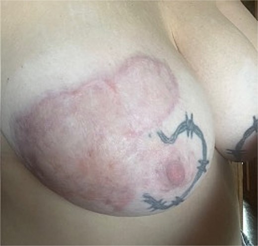
Over the next 48 h, the patient systemically improved; however, she developed rapidly progressive bruising over the lateral breast. Concerns at this stage were raised for necrotizing fasciitis. By this stage her breast milk cultures had returned with growth of Streptococcus pyogenes and methicillin-resistant Staphylococcus aureus (MRSA). Surgical consultant review was sought, with the impression that the patient had a Group A Streptococcus (GAS) erysipelas with haemorrhagic appearance (Fig. 1). This was the primary differential as by this time the patient was systemically well with minimal pain. Despite this, the patient was commenced on both GAS coverage and necrotizing fasciitis coverage. She remained well, without deterioration, and was discharged on Day 5 to hospital in the home for wound management and ongoing oral flucloxacillin.
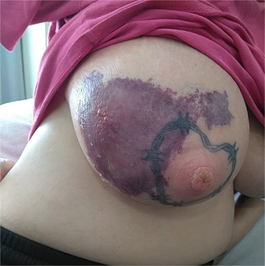
Haemorrhagic and bullous changes to right breast.
Approximately 2 weeks following discharge, the patient begun to develop dry necrosis. This had sloughy exudate superiorly and milk leakage from the wound (Fig. 2). She was taken to theatre for debridement and application of vacuum-assisted closure (VAC) dressing (Fig. 3). Following this she had three further returns for debridement, until the wound was clean, healthy and without ongoing milk leakage. Her wound continued to heal well, and she underwent elective debridement and skin grafting approximately 1 month following her initial operation. This healed well (Figs 4 and 5) and she was discharged from follow up. She remains well with no further episodes.
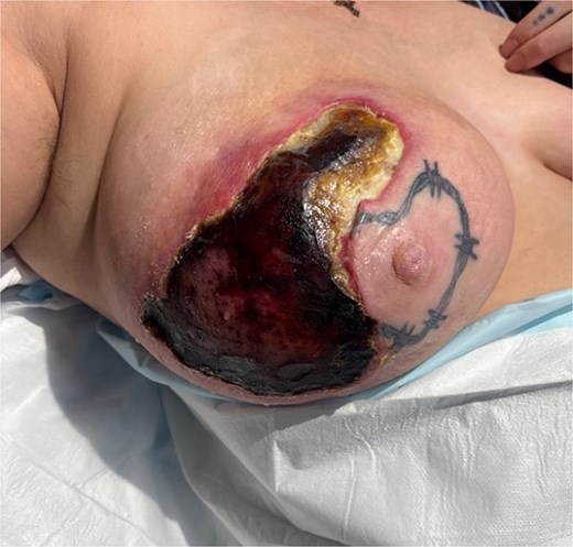
Two weeks post initial presentation, development of dry necrosis and milk leakage.
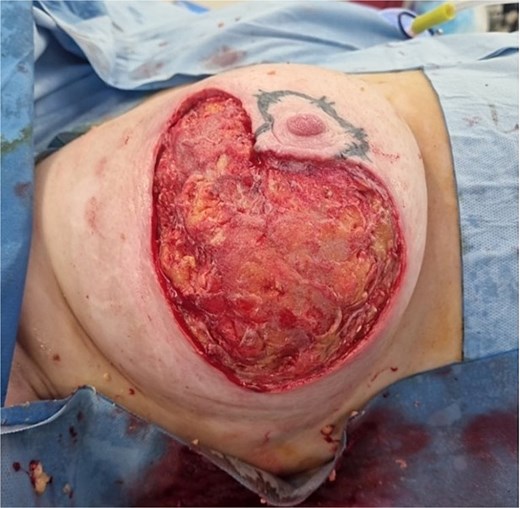
Intraoperatively, post initial debridement.
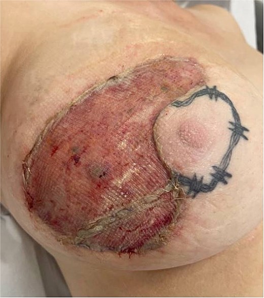
Initial postoperative skin graft.
Healed skin-graft, 6 months post grafting.
Discussion
Erysipelas refers to a skin infection that is localized to the dermis, most commonly affecting limbs and face [1]. It is characterized by well-demarcated, erythematous lesions and is often accompanied by fever [2]. Most commonly, the causative infective organism are Streptococci, and resolution occurs with simple treatment with penicillin, with which one can expect a rapid response [3]. However, there remains potential with erysipelas for development into more severe forms, such as with bullae or abscess formation, haemorrhagic transformation, or necrosis [4]. Where these do occur, they are most common in immunosuppressed patients, or those with pre-existing predisposing disease (e.g. lymphoedema, leg ulcers) [1].
Whilst a common infection more broadly, examples of breast erysipelas are scant, particularly with minimal descriptions of progression to necrosis. Few case reports describe breast erysipelas and typically are non-severe and with resolution following antibiotics [5]. The only clearly identified descriptions of similar severity to ours were found in a Russian article from 1993. This case series describes seven cases of postpartum breast necrosis caused by erysipelas, however, state that ‘as a rule’ it occurred in patients with concomitant heart or vessel disease [6]. Other case series describe breast necrosis, without preceding erysipelas and more commonly with preceding breast trauma [7]. Beyond this, most descriptions of erysipelas in relation to breast patients, are described in upper limbs following breast cancer treatment [8]. Occasionally, erysipelas-like changes have been observed over the breast themselves, though these have followed breast surgery [9]. This case appears to be the first documented case of an otherwise healthy female developing such severe breast erysipelas.
An important mimic of severe erysipelas is necrotizing fasciitis. This is prudent to exclude due to its time critical and life-threatening nature. Necrotizing fasciitis of the breast is a clinical emergency, also rare, with few cases described. Recent systematic reviews of existing literature suggest around 40 recorded cases in the last 20 years [10]. The difficulty in distinguishing diagnoses becomes evident in cases like ours; both have presence of pain and fevers, similar organisms [10], and overlap in appearance. However, one must consider the clinically distinguishing factors. Of note, a commonly described symptom of necrotizing fasciitis is ‘pain out of proportion,’ also described in breast cases [11]. Further, patients are typically systemically unwell, with this coinciding and worsening with deteriorating appearance of the affected tissue [11]. Conversely, our patient systematically improved by the time her breast developed haemorrhagic or bullous changes. Regardless, if consideration of necrotizing fasciitis is present, one must consider the potential benefit of empirical triple antibiotic coverage. Further, it is prudent to have senior surgical input to prevent a late or missed diagnosis of necrotizing fasciitis [10]. Distinguishing between these rare entities requires careful consideration.
Ultimately, such severe breast erysipelas in an otherwise healthy patient is extremely rare, and it is important to consider other life-threatening diagnoses. Key principles in management include early exclusion of necrotizing fasciitis, treatment with antibiotics, close monitoring and debridement of necrotic tissue as required. Once satisfied, reconstruction to best possible cosmetic outcome is ideal.
Conflict of interest statement
None declared.
Funding
None declared.
