


Abstract
Basal cell carcinomas are common facial malignancies with minimally invasive treatment approaches effective in the majority of cases. Recurrent aggressive lesions pose significant challenges and need wide local excision with major reconstruction. Geriatric patient with multiple comorbidities needs customized reconstructions to minimize morbidity. We present a case of longstanding invasive confluent basal cell carcinoma involving right cheek, ear lobule, and retroauricular area. The perforator plus variant of supraclavicular flap was successfully used for closure of the large defect.
Introduction
Basal cell carcinoma (BCC), a malignant epithelial neoplasm of the skin most often arises in area of chronic sun exposure [1]. Most (86%) of BCC are found in the head and neck region, where they are also most difficult to treat. Most lesions are managed by one of numerous minimally invasive options. Rarely, longstanding neglected cases may need wide local excision and major reconstruction. Restoration of function and esthetic considerations while minimizing morbidity, dictate the approach [2, 3]. However, obtaining these goals in large recurrent lesion of head and neck remains a more challenging task [4]. In accordance with the SCARE 2023 guidelines, we present a case of 82-year-old patient highlighting the use of a perforator-plus supraclavicular flap for reconstruction after the resection of extensive recurrent basal cell carcinoma of the face [5].
Case report
An 82-year-old female was referred to our outpatient department with a 5-year history of progressive nodular ulcerative swelling on the right side of her face. She had a history of similar lesions on right cheek, having undergone excision 13 years ago and re-excision for recurrence on the right cheek 10 years ago. The lesions were pruritic, with frequent serosanguinous discharge (Fig. 1). On examination, the lesions were nodular ulcerative with hyperpigmentation and no regional lymphadenopathy. Biopsy revealed multiple nests of basaloid cells with peripheral palisading, surrounded by a fibrous stroma, suggestive of basal cell carcinoma (BCC).
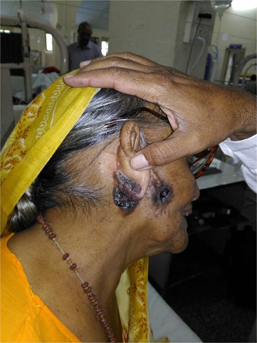
Nodular ulcerative lesion on the right cheek and ear lobule.
The lesion was excised with a 7 mm margin; resulting in a defect measuring 11 × 8 cm, extending across right cheek, ear lobule, and retroauricular area (Fig. 2).
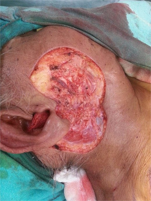
Extensive defect across the right cheek, ear lobule, and retroauricular area after lesion excision.
A supraclavicular perforator plus flap was planned to reconstruct the defect. Doppler ultrasound confirmed the presence of supraclavicular artery and the flap was raised subfascially from distal to proximal in the standard fashion. Once the vascular pedicle was reached, the flap inset was checked but found deficient. Therefore, the flap was extended proximally beyond the pedicle until it could be inset without tension. The overall flap dimension after elevation was 22 × 9 cm. The flap was not islanded instead it was retained as a random transposition flap with base intact. The flap was inset into the defect by incising the skin bridge, avoiding the need for de-epithelialization. The donor site was closed primarily and a single suction drain employed under the donor site closure and flap (Fig. 3a and b).
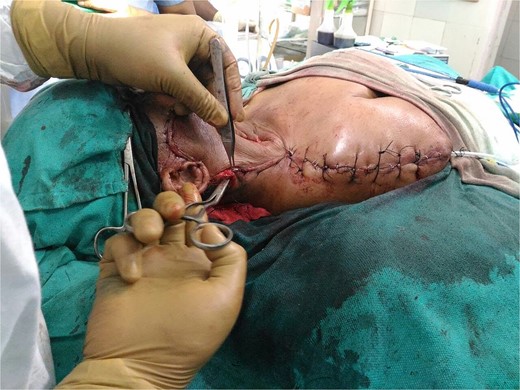
Flap inset into the defect with donor site closer and suction drain in place.
Postoperatively, the patient was positioned with her head elevated and neck neutral for 48 hours. Her recovery was uneventful and discharged on day 5. Histopathology revealed infiltrative BCC, Clark level V, stage pT2NxMx, with clear margins. On follow up at 2 weeks the wound was healthy and suture removal was done (Fig. 4) and on 12 months of follow-up, there were no sign of reccurrence.
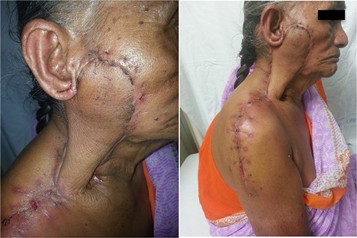
Healed surgical site at 15 days post-op.
Discussion
Our patient was an infirm 82-year-old female who had a long-standing lesion with multiple recurrences. A single-stage wide excision was performed, followed by flap reconstruction. Though numerous reconstructive techniques exist for smaller defects, large defects like the one in this case are best addressed with more advanced approaches. One has to rely on reconstructive golden principles to replace like with like. In this regard loco-regional flaps offer the best approaches with minimal morbidity.
The 11 × 8 cm defect was too extensive for local flaps, so we chose a regional supraclavicular fasciocutaneous flap due to its thin, pliable tissue, rapid elevation, and minimal donor site morbidity, making it an excellent option for reconstruction. However, a known drawback of this flap is necrosis, reported in up to 22% of cases with island flaps. This complication is often attributed to twisting of the vascular pedicle, leading to arterial insufficiency, or venous congestion.
Recent understanding of perforator anatomy has led to reclassifying the island flap as a supraclavicular perforator flap [6]. We believe that retaining the flap base provides an additional random arterial input to buttress the supply from the supraclavicular pedicle. The retained flap base also provides an additional route for venous and lymphatic outflow. This variation is a form of the perforator plus flap, which has been described as a flap retaining the main perforator as well as the flap base [7]. As can be seen from postoperative images, the flap does not show any evidence of ischemia, edema, or congestion. The retained base also prevents an acute twist of the vascular pedicle. De-epithelializing the base could compromise the subdermal vascular plexus, reducing arterial input, while tunneling may impair survival through compression and edema. We retained the flap base and inset it through an oblique neck incision bridging the defect and donor site. A disadvantage may be the minimal dog ear, which settles with compression and time with a good esthetic outcome. We feel that the perforator plus variant of the supraclavicular flap is safe, enhances the blood supply, and drainage leading to larger flap insets with decreased incidence of necrosis. Although this case demonstrates the feasibility of this approach with nearly 170-degree rotation, a series of cases would be necessary to confirm its advantages.
Conclusion
The perforator-plus supraclavicular flap can be a reliable and versatile option for reconstructing large defects in the head and neck following resection of recurrent BCC. By retaining the flap’s base, this technique enhances blood supply and minimizes the risk of necrosis, making it particularly suitable for elderly patients with comorbidities.
Acknowledgements
We dedicate this case report to the memory of Brig Sandeep Mehrotra, plastic surgeon par excellence, whose mentorship and guidance were pivotal in making this case report possible. His passion for surgery and commitment to excellence continue to inspire all who had the privilege of learning from him.
Author contributions
Conceptualization: AT, AS. Patient Management: AT, AS. Writing – original draft: AT, PL, AS. Writing – review & editing: AT, PL, AS, SP, AS. Visualization and Supervision: AT, AS.
Conflict of interest statement
There were no conflicts of interest among all authors.
Funding
This study received no funding.
Data availability
The details are available from corresponding author upon reasonable request.
Guarantor
Prajjwol Luitel.
Declaration
All the authors declare that the information provided here is accurate to the best of our knowledge.
Consent
Written informed consent was obtained from the patient for publication and any accompanying images. A copy of the written consent is available for review by the Editor-in-Chief of this journal on request.
