-
PDF
- Split View
-
Views
-
Cite
Cite
Marco Di Mitri, Greta Chiastra, Edoardo Collautti, Simone D’Antonio, Marina Buzzi, Cristian Bisanti, Annalisa Di Carmine, Vincenzo Catania, Michele Libri, Tommaso Gargano, Mario Lima, Platelet-rich plasma therapy for postoperative esophageal fistula in a pediatric patient, Journal of Surgical Case Reports, Volume 2024, Issue 5, May 2024, rjae350, https://doi.org/10.1093/jscr/rjae350
Close - Share Icon Share
Abstract
Postoperative management of esophagocutaneous fistulas in pediatric patients is challenging, often resulting in prolonged hospitalization and increased morbidity. Platelet-rich plasma (PRP) has emerged as a promising adjunctive treatment for such complications. We present the case of a 7-month-old infant who developed an esophago-cutaneous fistula following esophagocoloplasty for esophageal atresia type A. Despite initial conservative management, the fistula persisted, prompting the application of PRP gel derived from umbilical cord blood. After four applications of PRP, complete closure of the fistula was achieved, leading to both functional and aesthetic results. This case highlights the potential of PRP in managing refractory postoperative esophageal fistulas in pediatric patients and underscores the need for further research to optimize treatment protocols and validate its efficacy for this sort of complications
Introduction
Esophageal fistula following surgery represents a challenge in pediatric patients, often leading to prolonged hospital stay, increased morbidity, and potential mortality. These postoperative complications can arise from a variety of procedures, including esophageal atresia (EA) repair, antireflux surgery, esophagectomy, and trauma [1–4]. Despite the advancement in surgical techniques and perioperative care, the management of postoperative esophageal fistulas remains complex and requires innovative therapeutic strategies. Among these strategies, platelet-rich plasma (PRP) has emerged as a promising adjunctive treatment in several medical disciplines [5]. PRP is derived from the patient’s own blood or from a donor and contains a concentrated mix of platelets, growth factors, and other bioactive proteins that play key roles in tissue repair and wound healing [6]. Its ability to harness the regenerative potential of growth factors makes it an appealing option for addressing challenging clinical scenarios, such as postoperative complications. In this report, we present a case of an infant who developed an esophago-cutaneous fistula following esophagocoloplasty for EA type A. Through this case presentation, we aim to highlight the difficulties in managing postoperative esophageal fistulas in pediatric patients, focusing on the potential role of PRP.
Case report
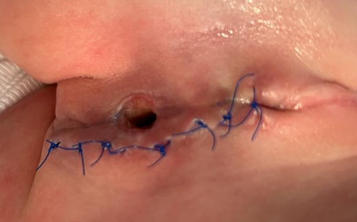
A newborn, female, born at full term (38 weeks of gestational age) after a regular pregnancy, except for the discovery of polyhydramnios and an absent gastric bubble, which led to suspicion of EA. The diagnosis (type A EA) was confirmed at birth by performing an X-ray scan and a laryngotracheoscopy which excluded the presence of trachea-esophageal fistula. The gap between the two esophageal stumps was of three vertebral bodies. Due to this gap, at the age of 2 months, the patient underwent the Foker’s technique of the extra-thoracic thoracoscopic-assisted elongation of the two esophageal stumps. After 10 days, the attempt to perform the esophageal anastomosis was unsuccessful, leading to a left cervical esophagostomy in preparation for esophageal substitution. At the age of 7 months, the infant underwent the esophagocoloplasty procedure. On the 14th postoperative day, the patient developed an esophago-cutaneous fistula above the proximal esophagus-colon anastomosis, resulting in copious saliva leakage (Fig. 1).

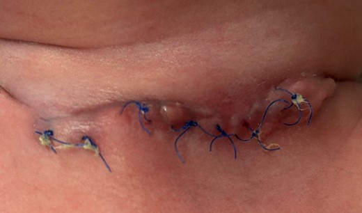
We initially pursued a conservative approach, which included total parenteral nutrition and daily advanced dressings of the fistula. After 2 months, even though the fistula appeared to have decreased in size, saliva leakage persisted (Fig. 2).

After 2 months of conservative management and advanced dressing.
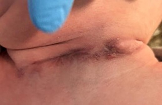
Due to the persistence of symptoms, we decided to use platelet-rich plasma gel (PRP-G), derived from umbilical cord blood (CB) and prepared at the Emilia Romagna Cord Blood Bank in S. Orsola Hospital (Bologna-Italy). We applied the PRP-G four times, renewing it every fourth day. Remarkably, by the third application, the fistula had completely closed. We chose to apply it one more time to optimize wound healing. Through the use of PRP, we obtained both the closure of the fistula and the development of a pleasing scar (Fig. 3).

Cord blood platelet gel preparation
After the mothers’ informed consent compilation, CB units were collected by trained midwives into plastic bags containing 25 ml of citrate–phosphate–dextrose anticoagulant, before and after placental delivery. After storage and transportation at monitored temperature %to the Emilia Romagna Cord Blood Bank in S. Orsola Hospital, the units were processed within 48 h of collection. Following two centrifugations and the removal of supernatant platelet-poor plasma (PPP), a platelet concentration between 800 × 109/ and 1200 × 109/l in PRP was obtained. The PRP units were transferred into a storage bag and cryopreserved in a mechanical freezer at a temperature below −40°C. Microbiological control was performed on PPP. To obtain a CB platelet gel, the PRP was thawed at 37°C in a water-bath and then activated with calcium gluconate (with or without thrombin) at 37°C inside an incubator. The platelet gel was then delivered to the pediatric surgery department for application.
Discussion
The case presented underscores the challenges associated with postoperative esophageal fistulas in pediatric patients, particularly following complex surgical procedures such as esophagocoloplasty. Despite advancements in surgical techniques, the development of esophageal fistulas remains a significant complication, often requiring innovative therapeutic strategies to achieve favorable outcomes [7]. PRP has turned out to be a promising adjunctive treatment for in our case, demonstrating its potential in promoting tissue repair and wound healing [5, 8]. PRP, which was derived from umbilical CB in this instance, contains a concentrated mix of platelets and growth factors that play a crucial role in the healing process [6, 9]. By harnessing the regenerative potential of these growth factors, PRP therapy offers a novel approach to address challenging clinical scenarios such as postoperative complications [10, 11]. In our case, the first-line conservative management failed to resolve the esophago-cutaneous fistula, highlighting the need for alternative therapeutic solutions [12]. The decision to use PRP-G was based on its known regenerative properties and its potential to stimulate tissue repair. Remarkably, after four applications of PRP-G, the fistula achieved complete closure, leading to both functional and aesthetic improvements. The successful closure of the fistula following PRP therapy underscores its potential utility in the management of refractory postoperative esophageal fistulas in pediatric patients. Further research, including larger-scale studies with controlled designs, is warranted to validate the efficacy and safety of PRP therapy in this patient population. Additionally, the optimal protocol for PRP application, involving timing, frequency and dosage, warrants further investigation. Standardization of PRP therapy protocols will facilitate its integration into clinical practice and enhance its reproducibility across different healthcare settings. In conclusion, our case highlights the potential of PRP therapy as a promising adjunctive treatment for postoperative esophageal fistulas in pediatric patients. By exploring innovative therapeutic strategies, such as PRP, we can improve the management of postoperative complications and ultimately enhance clinical outcomes in pediatric surgical care. Further research is needed to elucidate the optimal use of PRP and its long-term effects in this patient population.
Conflict of interest statement
The authors declare they have no competing interests.
Funding
No funding was received for conducting this study.
Data availability
Please contact author for data requests.
Ethics approval
All procedures performed in studies involving human participants were in accordance with the ethical standards of the institutional and/or national research committee and with the 1964 Helsinki Declaration and its later amendments or comparable ethical standards.
Informed consent
Informed consent was obtained from all subjects involved in the study.
Consent to publish
The participant has consented to the submission of the case report to the journal. The authors affirm that the participant provided informed consent for publication of the images in Figs 1–3.
References
- pathologic fistula
- colorectal surgery
- esophageal atresia
- esophageal fistula
- esthetics
- gel
- infant
- pediatrics
- postoperative care
- surgical procedures, operative
- urology
- morbidity
- plastic surgery specialty
- plastic surgery procedures
- pediatric surgery
- umbilical cord blood
- upper gastrointestinal tract series
- platelet rich plasma
- conservative treatment
- pediatric surgical procedures


