-
PDF
- Split View
-
Views
-
Cite
Cite
Brice Blum, Arthur D Grimes, Hannah L Carroll, Gregory R Stettler, Cecal volvulus secondary to mesodiverticular band, Journal of Surgical Case Reports, Volume 2024, Issue 5, May 2024, rjae296, https://doi.org/10.1093/jscr/rjae296
Close - Share Icon Share
Abstract
Meckel’s diverticula are one of the most common gastrointestinal anomalies, yet mesodiverticular bands are rare. The treatment of these bands commonly requires surgery. A healthy patient in his 20s presented to the emergency department with a 1 day history of acute onset abdominal pain. Computed tomography imaging was consistent with volvulus of the large intestine. In the operating room, the patient was noted to have a band between the ileal mesentery and tip of a Meckel’s diverticulum, consistent with a mesodivertiular band, through which cecum had volvulized. The patient underwent resection. The patient recovered without major complications. Mesodiverticular bands are rare, but may present as hemoperitoneum, small bowel obstruction, or volvulus. Pre-operative diagnosis of a mesodiverticular band is often difficult and they are most commonly diagnosed intraoperatively. Treatment should include surgery and may include simple lysis of the band, bowel resection, or more extensive resection if other pathology is present.
Introduction
A Meckel’s diverticulum is the most common congenital abnormality of the small bowel with incidence in the range 1%–4% of the population [1]. A mesodiverticular band is an obliterated remnant of the vitelline artery, which embryologically supplies blood to a Meckel’s diverticulum [2, 3]. It usually connects the tip of the Meckel’s diverticulum to the mesentery of the ileum [2]. It is the result of failure of involution of the omphalo-mesenteric ligament, which usually obliterates by the 10th week of gestation [3]. This rare entity has previously been noted to present as bowel obstruction or hemoperitoneum, more commonly in male patients than female patients [2, 4–6]. Anatomically, the mesodiverticular band creates a snare-like opening through which loops of bowel can herniate, become obstructed, and, in severe cases, become strangulated, making early diagnosis of this pathology very important [2]. Herein, we identify a rare case of mesodiverticular band creating a cecal volvulus with acute obstruction.
Case report
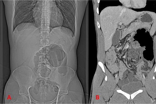
A healthy patient in his 20s, with no previous abdominal surgical history, presented to the emergency department with a 1 day history of acute onset abdominal pain. The patient presented with normal vital signs and normal labs. His physical exam was notable abdominal distention as well as significant lower abdominal tenderness and rebound, consistent with focal peritonitis. Computed tomography (CT) scan of the abdomen and pelvis was obtained which revealed volvulus of a segment of large intestine with radiographic findings concerning for ischemic changes (Fig. 1A and B). Imaging was concerning for a large bowel volvulus. As a result, it was recommended to proceed to the operating room for exploration given concern for ischemia.

Imaging showing findings concerning for volvulus of the large intestine; (A) plain film of abdomen; (B) coronal CT imaging.
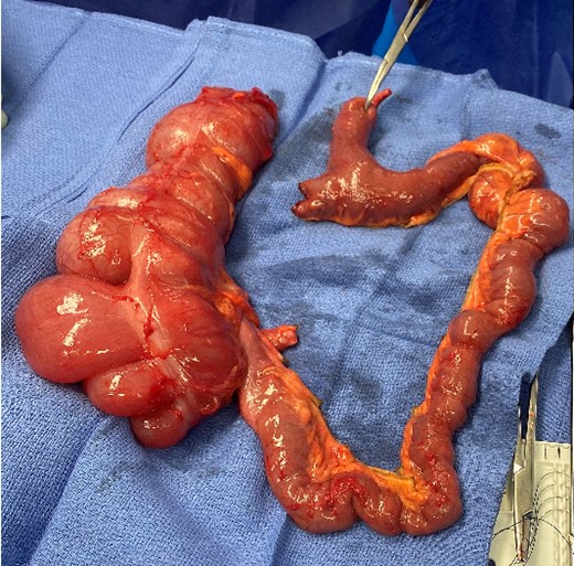
In the operating room, we proceeded to perform a midline laparotomy, choosing to forego attempts at a laparoscopic exploration given radiographic findings of colonic volvulus and peritonitis on exam. Upon entry, there was no evidence of peritoneal contamination. Exploration revealed a normal sigmoid colon. However, the cecum was volvulized under a band that connected from the tip of a Meckel’s diverticulum to the ileal mesentery, under which the redundant and ischemic cecum had volvulized. The diverticulum measured to be ~60 cm from the ileocecal valve. There was no other evidence of additional intraabdominal pathology. In the presence of strangulation, the decision was made to resect the redundant cecum as well as the Meckel’s diverticulum with associated mesodiverticular band (Fig. 2). The bowel was divided between GIA staplers with the mesentery divided with an energy device. A stapled side-to-side anastomosis utilizing the Barcelona technique was completed to restore intestinal continuity and the mesenteric defect was closed.

Surgical specimen of en bloc resection of redundant colon, small bowel, and Meckel’s diverticulum; instrument is identifying based of mesodiverticular band.
Postoperatively, the patient recovered well. The post-operative course was complicated by fever and small intraabdominal fluid collection treated successfully with antibiotic therapy. The patient was discharged on post-operative Day 8. At clinic follow-up 2 weeks following discharge, the patient was recovering well without complaints or other complications. Final pathology revealed cecum and small bowel with evidence of ischemic changes.
Discussion
Meckel’s diverticulum is one of the most common congenital anomalies of the gastrointestinal tract. A Meckel’s diverticulum is most often asymptomatic; however, it can cause a litany of complications including ulceration, hemorrhage, intussusception, obstruction, perforation, or can even be associated with fistulas and malignancy [7]. It can present symptomatically either in children or adults, but can also be identified incidentally during an operation or by cross-sectional imaging [3].
The blood supply to a Meckel’s diverticulum is from the vitelline artery. Along with the omphalo-mesenteric ligament, this structure usually involutes during gestation. Failure of this structure to involute and obliterate leads to a mesodiverticular band, the embryologic remnant of the vitelline artery and blood supply to a Meckel’s diverticulum [2, 4]. There have been few reports describing pathology associated with mesodiverticular bands, as it is a rare entity. However, cases that have been presented generally describe a presentation of small bowel obstruction and rarely hemoperitoneum. It is further recommended that if these bands are identified incidentally, during an operation for another cause, then they be lysed or excised to reduce the risk of future herniation or volvulus [2]. Even more rare than small bowel obstruction are descriptions of a cecal volvulus secondary to a mesodiverticular band [2].
As this entity is rare, diagnosis can often be a challenge [2, 4]. Most commonly, a mesodiverticular band is identified at the time of surgery, and as such should be addressed when identified. However, there are reports of radiographic identification of a mesodiverticular band [4]. In the largest systematic review of patients that had been identified with a mesodiverticular band (either at time of operation or postmortem), 95% of patients had undergone surgery. The one patient that did not undergo surgery ultimately was noted to have strangulated bowel secondary to a mesodiverticular band on postmortem examination. This underscores the importance of surgery in the treatment of this pathology. Options for surgery are lysis of band, diverticulectomy, small bowel resection, or, as in our case, en bloc resection of colon and diverticulum with intervening small bowel [2, 4].
Conclusion
Mesodiverticular bands are rare causes of intestinal pathology and can present as obstruction, hemoperitoneum, or volvulus. The presence of a symptomatic mesodiverticular band usually requires surgery to alleviate symptoms and potentially resect compromised bowel. Often times, these bands are not identified until abdominal exploration and the surgeon has several surgical interventions including lysis of band, diverticulectomy, and bowel resection at their disposal to treat this rare entity.
Author contributions
Brice Blum participated in the care of the patient, drafted, and critically revised the manuscript. Arthur D. Grimes and Hannah L. Carroll drafted, and critically revised the manuscript. Gregory R. Stettler participated in the care of the patient, drafted, and critically revised the manuscript.
Conflict of interest statement
None to declare.
Funding
This research received no specific grant from any funding agency, commercial, or not-for-profit sectors.
Patient consent
After review of ICMJE Protection of Research Participants, consent for publication was not obtained in this case as the manuscript was deidentified and provides appropriate patient anonymity.
References
- a band
- abdominal pain
- small bowel obstruction
- computed tomography
- bowel resection
- congenital anomaly of gastrointestinal tract
- emergency service, hospital
- hemoperitoneum
- intestine, large
- mesentery
- operating room
- surgical procedures, operative
- cecum
- diagnosis
- diagnostic imaging
- ileum
- meckel's diverticulum
- pathology
- cecal volvulus
- intestinal volvulus
- cytolysis


