-
PDF
- Split View
-
Views
-
Cite
Cite
Asama Rana, Zamaan Hooda, Sayali Kulkarni, Karmina Choi, An unusual case of gallstone ileus within the cecum and ascending colon: a case report, Journal of Surgical Case Reports, Volume 2023, Issue 6, June 2023, rjad327, https://doi.org/10.1093/jscr/rjad327
Close - Share Icon Share
Abstract
Gallstone ileus is a rare cause of intestinal obstruction. Due to long-standing inflammation of the gallbladder, fistulization can occur within nearby structures, most commonly to the duodenum or hepatic flexure of the colon. Through these fistulas, a stone can migrate and result in a small bowel obstruction or a large bowel obstruction. This case exemplifies the diagnosis and treatment of gallstone ileus, along with potential complications due to stone migration. Early recognition and treatment of gallstone ileus is important, as stone migration can lead to increased mortality with delayed diagnosis.
INTRODUCTION
Gallstone ileus is a rare etiology of mechanical bowel obstruction. It accounts for less than 5% of all intestinal obstructions with a higher frequency in elderly patients [1, 2]. Gallstone ileus occurs in 0.3–0.5% of patients with a history of cholelithiasis, with a strong predominance for women [3, 4]. The development of this disease is due to chronic inflammation of the gallbladder wall leading to dense adhesions around adjacent structures causing fistulization [5]. The most common sites of fistula formation are the duodenum (cholecystoduodenal fistula), the stomach (cholecystogastric fistula) and less commonly within the colon (cholecystocolonic fistula) [2, 6]. Stone migration starts in the gallbladder and most commonly flows through the cholecystoduodenal fistula then ends up at the terminal ileum [1]. Stones greater than 2 cm will be unable to flow past the ileocecal valve leading to a small bowel obstruction [1, 7]. Much rarer, a stone can flow through the cholecystocolonic fistula and reach the narrow walls of the sigmoid colon ultimately leading to a large bowel obstruction [7, 8].
We present a case of a small bowel obstruction secondary to gallstone ileus with subsequent migration of the stone to the cecum and ascending colon.
CASE REPORT
This is an 84-year-old woman who presented to the emergency department with acute onset abdominal pain. She described her pain as colicky and diffused for 3 weeks, with worsening pain in the periumbilical region and left lower quadrant. Her pain was associated with nausea and multiple episodes of vomiting, and her last bowel movement was 2 days prior to admission.
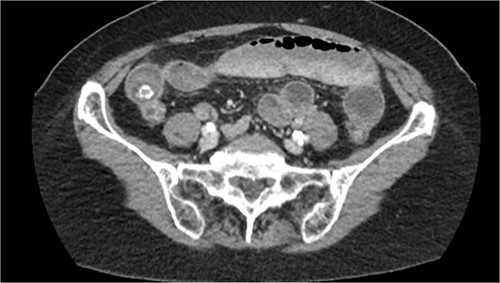
On physical exam, the abdomen was notable for diffuse periumbilical tenderness without guarding, rigidity or any palpable masses. Laboratory studies were all within normal limits. CT scan of the abdomen and pelvis with IV contrast demonstrated a small bowel obstruction, secondary to gallstone ileus with a severely inflamed gallbladder and fistulization to the duodenum and hepatic flexure of the colon (see Fig. 1). Two gallstones were identified within the ileum, 2 and 2.5 cm in size. Surgery was consulted, and a small bowel enterotomy was planned to retrieve the obstructing stone.

A diagnostic laparoscopy was performed. The small bowel was run from the ileocecal valve to the ligament of Treitz. The two gallstones previously visualized on CT were not identified within the small bowel. There were dense omental adhesions seen over the gallbladder and hepatic flexure. The omentum was taken down partially in order to visualize the gallbladder, which was severely inflamed and densely adherent to the adjacent duodenal bulb and hepatic flexure. In order to localize the obstructing gallstones, a laparoscopic hand-assist GelPort was placed within the infraumbilical incision. The entirety of the small bowel, including the duodenum, was exteriorized and examined, with no stones palpated. However, regions of erythema and submucosal hematomas were noted along the length of the small bowel suggesting mucosal injury due to the transiting gallstones. The colon was then examined. The 2-cm gallstone was palpated in the cecum, and the 2.5-cm stone was palpated in the ascending colon. The stones were attempted to be milked distally to retrieve transanally; however, they were not able to pass through the sigmoid, which was narrow with diffuse diverticulosis. The stones were then milked proximally into the mid-transverse colon. A 2-cm colotomy was made in the mid-transverse colon, and the two stones were retrieved. The patient tolerated the surgery well with no complications, and was extubated and transferred to the post anesthesia care unit in stable condition.
Postoperatively, the patient tolerated oral feeds, and was able to pass flatus and have normal bowel movements. She was discharged with oral antibiotics for ongoing cholecystitis on postoperative day 4 with plans for cholecystectomy in the near future.
DISCUSSION
Gallstone ileus manifests in 0.3–0.5% of patients with previously diagnosed cholelithiasis and makes up less than 5% of all cases of intestinal obstruction, with a predilection for women [1, 3, 4].
Given the nature and nonspecific symptoms of gallstone ileus, it is challenging to make a clinical diagnosis without additional diagnostic tools. The usage of CT scans, endoscopic studies and ultrasound can lead to early detection. Visualization of a fistula tract from the gallbladder can be determined with such diagnostic modalities and assists the surgeon in development of a thorough treatment plan. Failure to identify a cholecystoenteric fistula preoperatively would lead to the surgeon performing a long and complicated procedure [9, 10]. Given the extensive inflammation that leads to fistulization, an operation at such a site would lead to increased chances of bleeding, perforation and increased susceptibility to infections due to contamination of the peritoneum with intestinal contents [11].
The mainstay of treatment for gallstone ileus is an enterolithotomy to relieve the obstruction point. This is done by making a longitudinal enterotomy and then milking the stone out of that incision site, followed by closure of bowel with a transverse suture [3]. However, this only treats the current issue, when the underlying cause of disease is the cholecystoenteric fistula. Treatment of both conditions in a single operation raises controversy given the immense risks associated with correction of the fistulized tracts, as mentioned above [11, 12]. In a systematic review of 1001 cases, Reisner et al. found an association with decreased mortality when performing an enterolithotomy alone versus an enterolithotomy with correction of the fistulas [1]. In the case of our operation, the decision was made to not correct the two fistulas given the severe inflammation surrounding the gallbladder wall. The risks of removal outweighed the benefits. Instead, we focused the treatment plan on stone retrieval. We attempted to stay minimally invasive and milk the stone to the rectum to retrieve it transanally. However, the stone could not pass through the sigmoid given its narrow walls and diffuse diverticulosis. We then performed an enterolithotomy at the mid-transverse colon and obtained both stones through the incision site. Thorough search for any remaining stones in the small bowel and large bowel is necessary in order to identify another source of obstruction [2].
CONFLICT OF INTEREST STATEMENT
None declared.
FUNDING
This research did not receive any specific grants from funding agencies in the public, commercial, or not-for-profit sectors.


