-
PDF
- Split View
-
Views
-
Cite
Cite
Kentaro Shinohara, Masaoki Hattori, Keiya Aono, Akihiro Hirata, Ryogo Ito, Motoi Yoshihara, Internal hernia in the medial inguinal fossa with a concurrent indirect inguinal hernia: a case report, Journal of Surgical Case Reports, Volume 2023, Issue 4, April 2023, rjad191, https://doi.org/10.1093/jscr/rjad191
Close - Share Icon Share
Abstract
A 51-year-old male was presented with abdominal pain and vomiting. Contrast-enhanced computed tomography showed a dilated small intestine with a sac-like appearance in the right lower abdomen. An internal hernia in the inguinal area was found during emergency laparoscopic exploration. The incarcerated small intestine was gently reduced, and the internal hernia sac was located in the right medial inguinal fossa. An indirect inguinal hernia was identified just behind the dissected internal hernia sac. The internal hernia sac was resected, and the indirect inguinal hernia was repaired through a transabdominal preperitoneal approach. The patient had no recurrence of hernia within the 20 months of follow-up after the surgery. This is the first case of an internal hernia in the medial inguinal fossa with a concurrent indirect inguinal hernia. The topographical relationship of these two hernias suggested that an indirect inguinal hernia may cause an internal hernia in the medial inguinal fossa.
INTRODUCTION
Although an internal hernia in the inguinal area rarely occurs, a few cases have been reported as internal hernias arising in the supravesical fossa [1, 2], a triangular area bounded by the medial umbilical ligament (MUL) and the median umbilical fold. The lateral space of the supravesical fossa is called the medial inguinal fossa, which lies between the MUL and inferior epigastric artery (IEA) [3]. While an external hernia in the medial inguinal fossa often develops as a direct inguinal hernia [4], an internal hernia in this area has never been reported.
We describe the first case of an internal hernia in the medial inguinal fossa concurrently identified with an indirect inguinal hernia. A key finding of the present case was that an indirect inguinal hernia may cause an internal hernia in the medial inguinal fossa.
CASE REPORT
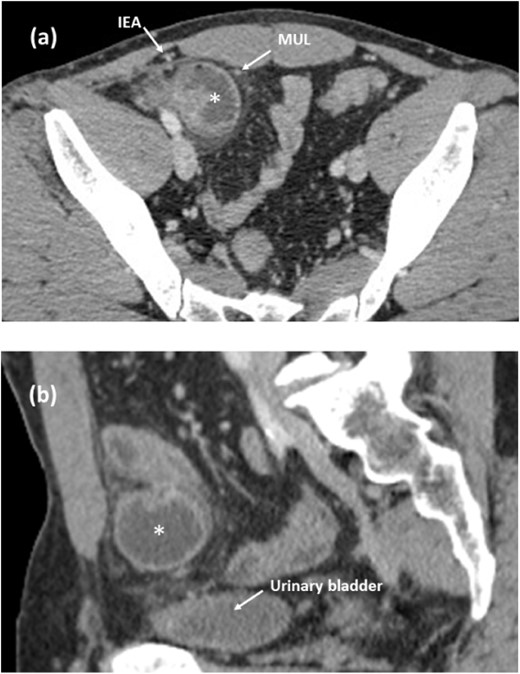
A 51-year-old man was presented with abdominal pain and vomiting. Contrast-enhanced computed tomography (CT) showed a dilated small intestine with a sac-like appearance in the right lower abdomen. The herniated small intestine was located between the MUL and the IEA (Fig. 1a) and did not compress the urinary bladder wall (Fig. 1b). The dilated inguinal canal was observed with extrusion of the extraperitoneal fat.

Contrast-enhanced CT images of the internal hernia in the medial inguinal fossa. (a, axial image) The herniated small intestine (asterisk) is located between the MUL and the IEA. (b, sagittal image) The herniated small intestine is apart from the urinary bladder. MUL, medial umbilical ligament. IEA, inferior epigastric artery.
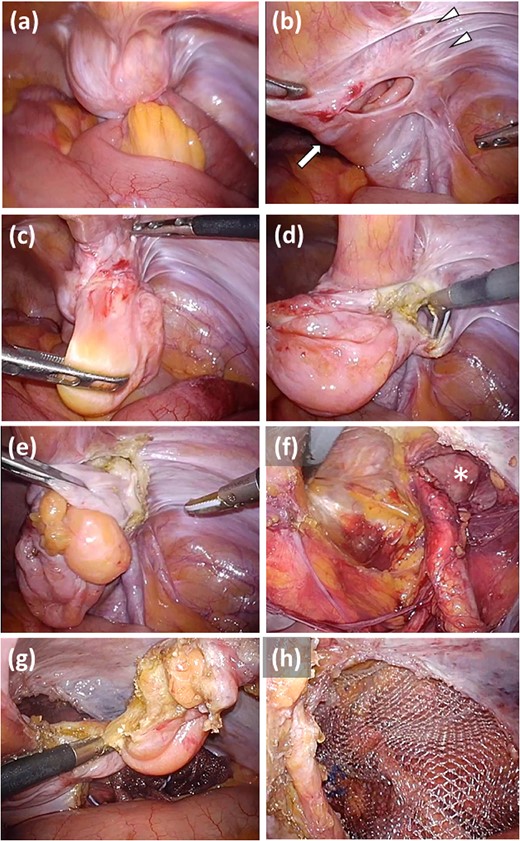
Emergency laparoscopic exploration was performed for acute small-bowel obstruction because of an internal Intraoperative findings showed that the small intestine was hernia. incarcerated in the preperitoneal fat around the right medial ligament (Fig. 2a). The herniated small intestine was gently reduced and had no evidence of strangulation. The hernia defect was located in the medial inguinal fossa, lateral to the right MUL (Fig. 2b). The internal hernia sac was adhered to the abdominal wall (Fig. 2b). The hernia sac was inverted and dissected from the lateral side to further explore the etiology of the hernia (Fig. 2c). Another peritoneum was identified just behind the dissected internal hernia sac (Fig. 2d), and it was found to be the sac of an indirect hernia (Fig. 2e and f). The residual peritoneum comprising the internal hernia sac was resected and removed (Fig. 2g). The indirect hernia was repaired via a transabdominal preperitoneal approach using a 3D Max® mesh (Bard-Davol, Warwick, USA) (Fig. 2h). The patient’s postoperative course was uneventful, and he had no recurrence of hernia within 20 months of follow-up after the surgery.

Intraoperative findings of the hernia repair. (a) The small intestine is incarcerated in the hernia sac. (b) The internal hernia sac located lateral to the right MUL (arrow) is adhered to the abdominal wall (arrowhead). (c) The internal hernia sac is inverted. (d) Another peritoneum is identified just behind the dissected internal hernia sac. (e) The peritoneum is the sac of an indirect hernia and is inverted. (f) Peritoneal dissection is performed to repair the indirect hernia (asterisk). (g) The internal hernia sac is resected. (h) The indirect hernia is repaired by using a transabdominal preperitoneal approach. MUL, medial umbilical ligament.
DISCUSSION
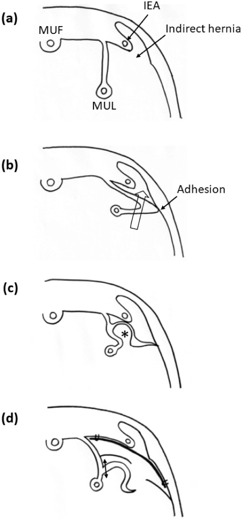
The present patient is the first case of an internal hernia in the medial inguinal fossa. The highlight of the present case is that a concurrent indirect hernia may have induced an internal hernia in the medial inguinal fossa. Laparoscopic exploration showed two pieces of evidence to support this hypothesis. One is the adhesion between the internal hernia sac and the abdominal wall lateral to the internal inguinal ring (Fig. 2b), and the other is that the indirect hernia sac was identified just behind the internal hernia sac (Fig. 2d). The presumed process of the development of the internal hernia in the medial inguinal fossa is as follows. An indirect inguinal hernia was first formed (Fig. 3a). The MUL adhered to the abdominal wall, and the adhered peritoneum was drawn into the indirect hernia cavity (Fig. 3b). The stretched peritoneum of the MUL ultimately provided the recess that incarcerated the small intestine (Fig. 3c).

Schematic illustration of the development of interna hernia in the medial inguinal fossa. (a) An indirect hernia is originally formed. (b) The MUL is adhered to the abdominal wall, and the adhered peritoneum is drawn into the indirect hernia cavity (arrow). (c) The stretched peritoneum of the MUL provides the recess that induces the internal hernia (asterisk). (d) Mesh repair is performed for the indirect hernia, and the internal hernia sac is resected (double arrow). MUF, median umbilical fold. MUL, medial umbilical ligament. IEA, inferior epigastric artery.
The present patient did not undergo simple closure of the hernia sac. Instead, we performed hernia sac resection and mesh repair for the indirect hernia detected just behind the internal hernia sac (Fig. 3d). Under the assumption that the indirect hernia caused the internal hernia in the medial inguinal fossa, simple closure of the internal hernia sac would have been insufficient, so mesh repair would be the optimal procedure to eliminate the primary factor inducing the internal hernia.
An internal supravesical hernia is another internal hernia arising in the supravesical fossa, and the etiology of this rare hernia is not yet known [1]. Although the internal hernia observed in the present patient did not meet the criteria of an internal supravesical hernia, the locations of these two internal hernias are adjacent to each other; therefore, these internal hernias may have the same etiology. In fact, reports show that ~25% of internal supravesical hernias occur concurrently with an inguinal hernia [1, 5]. Meticulous observation of the topographical relationship between the internal hernia sac and the concurrent inguinal hernia is key to clarifying the mechanism of internal supravesical hernia.
Laparoscopic exploration has been increasingly employed for an internal hernia [6, 7]. The present patient underwent laparoscopic surgery. Laparoscopic observation was useful for identifying the adhesion of the peritoneum, and reviewing the recorded video ensured the mechanism of the internal hernia in the present patient. Furthermore, simultaneous mesh repair of the indirect hernia was only feasible by using a laparoscopic approach. Although surgery for an internal hernia is generally performed in the emergency setting, using a laparoscopic approach would be effective in elucidating the mechanism of an internal hernia in the inguinal area.
In conclusion, the first reported internal hernia in the medial inguinal fossa may be caused by a concurrent indirect inguinal hernia. Laparoscopic observation helped elucidate the mechanism of this rare condition.
CONFLICT OF INTEREST STATEMENT
None declared.
FUNDING
None.


