-
PDF
- Split View
-
Views
-
Cite
Cite
Morimoto Shota, Toshiya Tachibana, Tomoya Iseki, Avulsion fracture of the calcaneal tuberosity treated with novel surgical technique using the combination of the side-locking loop suture technique and ring pins: a case report, Journal of Surgical Case Reports, Volume 2023, Issue 4, April 2023, rjad173, https://doi.org/10.1093/jscr/rjad173
Close - Share Icon Share
Abstract
A 78-year-old woman complained of right heel pain when tripping during walking. Radiographs revealed an avulsion fracture of the calcaneal tuberosity. Because the bone fragment was displaced, a novel osteosynthesis using the side-locking loop suture (SLLS) and ring pins was performed. At 3 months after the surgery, non-contrast computed tomography revealed complete bone union. At 2 years postoperatively, she had no symptoms and dysfunctions. Here, we describe a case of avulsion fracture of the calcaneal tuberosity in a 79-year-old female who treated with a novel surgical technique using combination the SLLS technique and ring pins. This surgical technique may be a useful option in the treatment for avulsion fractures of the calcaneal tuberosity.
INTRODUCTION
The avulsion fractures often occur by the traction force of the gastrocnemius–soleus complex because of minor trauma such as tripping because the insertion of the Achilles tendon is anatomically located in the calcaneal tuberosity, and which receives the tensile force of the gastrocnemius–soleus complex [1–3].
In general, surgical treatment is applied for the avulsion fractures with displacement [4, 5]. Various surgical procedures including lag screw fixation, tension band wiring, suture anchor technique and pullout technique have been reported [1, 5–11]. However, these procedures may be unable to fix the bone fragments rigidly enough to resist the tensile forces of the gastrocnemius–soleus complex because the bone fragments are often small and bone qualities of the calcaneus are poor [1, 9, 11]. Also, these procedures may lead to soft tissue complications and irritation [5, 11].
The side-locking loop suture (SLLS) technique has been reported as a surgical procedure for Achilles tendon ruptures [12]. Recently, surgical procedures using the SLLS technique for avulsion fractures of the calcaneal tuberosity have been reported [13, 14]. These surgical procedures can fix bone fragments rigidly enough to resist the tensile force of the gastrocnemius–soleus complex, whereas they require complicated processes when fixing bone fragments.
In this report, we describe an avulsion fracture of the calcaneal tuberosity in a 79-year-old female who treated with a novel surgical technique using the combination of the SLLS technique and ring pins. Written informed consent was obtained from the patient before publishing this report.
CASE REPORT
A 78-year-old woman complained of right heel pain when tripping during walking and visited to our hospital. She had medical histories such as arrhythmia, hypertension and hyperlipidemia. Physical examination revealed swelling, subcutaneous bleeding and tenderness on the posterior aspect of her heel and Thompson test was positive. Plain radiographs of the right ankle showed an avulsion fracture of the calcaneal tuberosity (Fig. 1). Non-contrast computed tomography (CT) demonstrated that the bone fragment was displaced. Based on medical histories, physical examination and image findings, we diagnosed an avulsion fracture of the calcaneal tuberosity and applied surgical treatment for the patient.
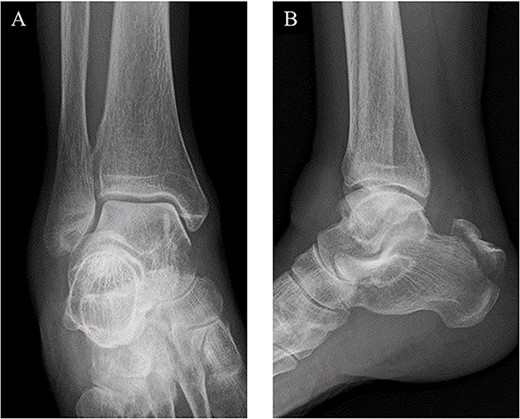
Plain radiographs of the right ankle showed no abnormal findings in (A) an anteroposterior view, but an avulsion fracture of the calcaneal tuberosity (B) a lateral view.
The surgery was performed under general anesthesia in the prone position with an air tourniquet and a fluoroscopy. First, an ~6 cm straight incision on the skin just above the calcaneal tuberosity was placed, and the fracture site was confirmed. After removing inclusions such as hematoma, the distal part of the Achilles tendon was sutured by the SLLS technique using USP 5 braided polyethylene and polyester suture thread (FiberWire; Arthrex Co., Naples, FL, USA) (Fig. 2A). Next, two 1.6-mm diameter ring pins (Tension Band Pin System; Acumed, Hillsboro, OR, USA) were parallelly inserted from the distal posterior part to the anterior part of the calcaneus body under a fluoroscopy (Fig. 2B). The bone fragment was anatomically reduced by pulling both of the suture ends, and the suture ends were passed through the rings of the ring pins from inside and outside, respectively. Then, the suture threads were crossed above the calcaneal tuberosity and were tied on the ventral side of the Achilles tendon (Fig. 2C). Finally, the wound was sutured, and the surgery was concluded (Fig. 3).
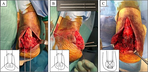
(A) The distal part of the Achilles tendon was sutured by SLLS technique using USP 5 braided polyethylene and polyester suture thread. (B) Next, two 1.6-mm diameter ring pins were parallelly inserted from the distal posterior part to the anterior part of the calcaneus body. (C) The bone fragment was anatomically reduced, and the suture ends were passed through the rings from inside and outside, respectively. The suture threads were crossed above the calcaneal tuberosity and were tied on the ventral side of the Achilles tendon.
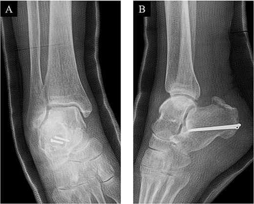
Postoperative (A) anteroposterior and (B) lateral ankle radiographs. The bone fragment was reduced and fixed using the combination of SLLS technique and ring pins.
Postoperatively, a non-weightbearing short leg cast was applied for 2 weeks, and when the cast was removed, active and passive ranges of motion exercises of the ankle were started. Partial-weightbearing was allowed at 2 weeks postoperatively, and full-weightbearing at 6 weeks postoperatively. At 3 months after the surgery, non-contrast CT showed complete bone union (Fig. 4). At 2 years after the surgery, she had no symptoms and dysfunctions (Fig. 5).
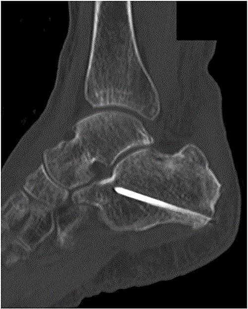
At 3 months after the surgery, non-contrast CT showed complete bone union in a sagittal view.
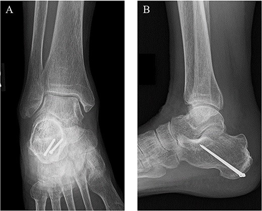
At 2 years after the surgery, plain radiographs of the right ankle showed complete bone union without the deformity of the fracture site and the backing out of the implants in (A) anteroposterior and (B) lateral views.
DISCUSSION
Various surgical procedures for avulsion fractures of the calcaneal tuberosity have been reported [1, 5–11]. Lag screw fixation is a most common surgical procedure [13, 14]. However, this procedure is difficult to fix bone fragments rigidly enough to resist the tensile force of the gastrocnemius–soleus complex when bone fragments are small and osteoporotic [1, 7, 8]. Squires et al. [5] introduced an oblique lateral tension band wiring fixation technique. This procedure is very bulky, and so may lead to soft tissue complications and irritation. Suture anchor technique be able to fix the bone fragments even if they are small, but the suture anchor insertion site is unable to resist the pullout force of the gastrocnemius–soleus complex [10]. Pullout technique was described by Banerjee et al. [11]. This technique be able to fix the bone fragment enough to resist the tensile force. However, it has a disadvantage that plantar incision is required and may lead to a painful scar.
Recently, surgical procedures using the SLLS technique for avulsion fractures of the calcaneal tuberosity have been reported [13, 14]. Miyamoto et al. [13] introduced a procedure using combination lag screw fixation and the SLLS technique. They treated two cases and reported this procedure fix the bone fragment rigidly and provided good surgical outcomes. However, this procedure requires complicated processes when tying and fixing the bone fragment. Wakatsuki et al. [14] treated one case by pullout fixation through the calcaneal bone tunnels using the SLLS technique. It has been reported that the procedure fixes the bone fragment rigidly and enable the early rehabilitation protocol. However, there is a disadvantage that plantar incision is needed in these procedures when fixing the bone fragment.
The ring pin is an interlocking system used in tension band wiring technique for patellar and olecranon fractures recently [15]. This implant has a small ring at the proximal end for passing the wire, and the proximal part is designed a low profile. Previous reports demonstrated that the use of the ring pins has advantages such as lower rates of implant irritation and preventing the backing out of the implants compared with the use of the Kirchner wires in tension band wiring technique for patellar and olecranon fractures [15].
In this report, we described an avulsion fracture of the calcaneal tuberosity in a 79-year-old female who treated by a novel surgical technique with the SLLS technique and ring pins. This surgical technique may be a useful option in the treatment for avulsion fractures of the calcaneal tuberosity.
CONFLICT OF INTEREST STATEMENT
None declared.
FUNDING
No funding.


