-
PDF
- Split View
-
Views
-
Cite
Cite
Moatasem Hussein Al-janabi, Ali Allan, Rabab Salloum, Incidental ectopic adrenal cortical tissue in the descending mesocolon in an elderly female: a first case report in the literature, Journal of Surgical Case Reports, Volume 2023, Issue 2, February 2023, rjad067, https://doi.org/10.1093/jscr/rjad067
Close - Share Icon Share
Abstract
Ectopic adrenal tissue (EAT) is an extremely unusual finding. The most common site is the genitourinary tract and pelvis, and more frequently in males than females. In our report, we discuss an ectopic adrenal cortical tissue detected in the descending mesocolon in an elderly female. To the best of our knowledge, this case is the first report in the English literature.
INTRODUCTION
Ectopic adrenal cortex tissue, also known as heterotopic adrenal tissue or adrenal rests, is an extremely unusual finding [1]. It is often discovered in the genitourinary system during early childhood and is considered to be found in less than 1% of adults, most commonly males [1, 2]. We report an exceptional finding of adrenal cortical rest located in the wall of descending colon, in a 57-year-old female noted by accident during the histopathological evaluation.
CASE PRESENTATION
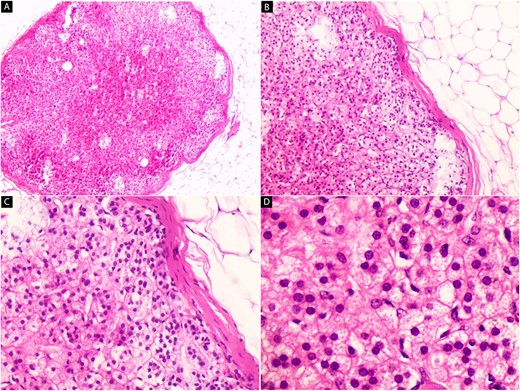
A 57-year-old female patient was referred to our hospital for a descending colectomy due to a previously diagnosed adenocarcinoma (signet ring type) and confirmed by histopathological examination. The abdominal examination was unremarkable. Routine blood tests were within normal limits. Descending colectomy is performed and the specimen was sent for histopathological examination. The tumor was submitted in five sections. During the dissection of the mesentery to find metastatic lymph nodes, a well-circumscribed small yellow nodule with a diameter of 0.5 cm was observed and placed in a separate cassette. One section from the tissue block was obtained for histologic study. On microscopic evaluation with hematoxylin and eosin staining, the nodule was composed of three layers of the adrenal cortex, outer zona glomerulosa an inner zona reticularis and, between them, the zona fasciculata (Fig. 1). No adrenal medullary tissue was observed. Macroscopic and histological features of this nodule are consistent with ectopic adrenal tissue (EAT) in the descending mesocolon.

Hematoxylin and eosin-stain (A–D). Microscopic images of the ectopic adrenal cortical nodule. (A) The low-power magnification shows a well-defined encapsulated nodule with adipose tissue around it (x40). (B) The nodule is composed of three adrenal gland cortical layers, zona glomerulosa (beneath the capsule), zona fasciculata (middle layer) and zona reticularis (innermost layer) (x100). (C) Small clusters and cords of polygonal cells with distinct cellular borders with lipid vacuoles are seen (x200). (D) The nuclei are round and located in the center of the cells (x400).
DISCUSSION
EAT, a rare entity, is often detected incidentally during surgery or pathological evaluation [1]. Ectopic adrenal rest is first discovered by Morgagni in 1740 [2]. EAT occurs in up to 50% of neonates, and <1% of adults. [2, 3]. Adrenal remnant tissue is more common in male than female children [1–3]. It is a benign lesion and mainly found in the genitourinary system, kidney, retroperitoneal fat, and rarely in the ovary, testis, spermatic cord, liver, transverse colon and gastric wall [1–3]. To the best of our knowledge, this case is the first report of EAT in the mesocolon in an elderly woman in the English literature, where a case report was described at this site, but in a newborn (Bal A,2009) [4]. Abnormal migration of mesoderm during the embryonic development of adrenal glands leads to dislocation of the adrenal tissue [3]. EAT is clinically silent, and a diagnosis can be made only by histopathological examination, usually as an incidental finding, as in this case [1, 3]. EAT is usually a single golden yellow nodule measuring 1 to 5 mm upon gross examination. Microscopically, it often consists of the three adrenal gland cortical layers, zona glomerulosa, zona fasciculata and zona reticularis, like in our case. Nevertheless, parts of the medulla can be found in some cases [5]. The diagnosis of heterotopic adrenal tissue should be kept in mind when a small yellowish nodule is observed in an extraordinary region during surgery or pathological evaluation [1, 6]. However, it is important to identify EAT and resect it because it may undergo the same pathological abnormalities, like hyperplasia and potential malignant neoplasm, that occur in the normal adrenal gland [1, 5, 7]. Consequently, surgeons should recognise this unusual finding, surgically remove it and send it for histopathological evaluation [1, 2, 7].
CONCLUSION
EAT is an extremely rare entity, particularly in the wall of the large intestine, which has no clinical significance in most cases. However, although EAT is an exceptional finding, it should be examined by pathologists to rule out potential malignancy.
CONFLICT OF INTEREST STATEMENT
None declared.
FUNDING
None.
CONSENT
Written consent was obtained.
GUARANTOR
Moatasem Hussein Al-janabi.


