-
PDF
- Split View
-
Views
-
Cite
Cite
Faranak Olamaeian, Mahdi Saberi Pirouz, Fatemeh Sheibani, Ali Tayebi, Amyand’s hernia: non incarcerated, inflamed appendix in inguinal sac case report, Journal of Surgical Case Reports, Volume 2022, Issue 9, September 2022, rjac382, https://doi.org/10.1093/jscr/rjac382
Close - Share Icon Share
Abstract
Inguinal hernia is described as protrusion of abdominal structures into inguinal canal, such as intestinal loop and abdominal fascia. Appendix rarely bulges into inguinal canal which is called Amyand’s hernia. A 55-year-old diabetic male presented to an outpatient clinic with right inguinal bulging since 2 years ago which was non-tender, without erythema and became non reducible since 2 days ago. Also bulging worsened by physical activity. The patient went through operation and an inflamed appendix was found stuck in hernia sac. Non incarcerated inguinal hernia can be diagnosed with physical examination and there is no need of further imaging which makes it hard to diagnose the nature of protrusion. Amyand’s hernia usually presents with pain due to appendicitis which mimics incarcerated hernia and makes it easier to suspect the etiology and request for further investigation. However in this case, pain was suppressed and this patient was candidate for elective inguinal herniotomy.
INTRODUCTION
Inguinal hernia is defined as protrusion of internal structure and fascia through inguinal canal. It is a high prevalent disorder in general population [1]. Omentum and intestinal loops are common structures that get stuck in inguinal canal. Also appendicitis, like inguinal hernia, is a very common and usually simple general surgery [2]. However, combination of appendicitis and incarceration of appendix into inguinal hernia is very rare (<1% of cases). According to the estimates, an inflamed perforated appendix is detected in the incarcerated inguinal hernia sac in only 0.13% of all operated patients [3, 4]. Amyand’s hernia is the protrusion of appendix in inguinal sac. It is named after English surgeon Claudius Amyand who performed the first appendectomy with perforated appendix within right inguinal cavity [5]. An appendix incarcerated in the right inguinal hernia sac is only found in extremely rare cases. However, cases vary in ages from neonates to elderly [6]. Inguinal hernia diagnosis is routinely based on clinical features and is not dependent on para clinical findings. Therefore, it is almost impossible to find out exact organ caught in hernia sac before surgery. Lichtenstein is the preferred technique for inguinal hernia. This tension free with prosthetic mesh operation is better tolerated by patient with lower risk of recurrence and hospitalization [7, 8]. If appendicitis is proved (type 2), appendectomy can be done during inguinal herniotomy. Also using of prosthetic materials in this type is not recommended and it is suggested to apply Bassini technique instead [9].
CASE PRESENTATION
A 55-year-old diabetic male patient with mental illness lives in a welfare center, and had a history of herniorraphy in his childhood presented to the outpatient surgical clinic at Firouz-Abadi Hospital with a history of right inguinal hernia from 2 years ago which became non reducible and a bit painful since 2 days before consultation. Also the bulging worsened by physical efforts.
At physical examination, bulging was seen in the right inguinal region. The approximate size of inguinal bulging was about 4–5 cm and the protrusion became more prominent by Valsalva maneuver. Patient had non abdominal pain and there were no evidence of pathological problem. However, the history was not completely reliable due to patient’s mental disorder.
Our diagnosis was inguinal hernia without incarceration and strangulation and the patient was candidate for elective inguinal hernia surgery. Preoperative lab tests were completely normal. Also abdominal ultrasound imaging reported no pathological finding except right inguinal hernia without incarceration. Further investigation was unnecessary. He was admitted to surgery department and prepared for right inguinal repair.
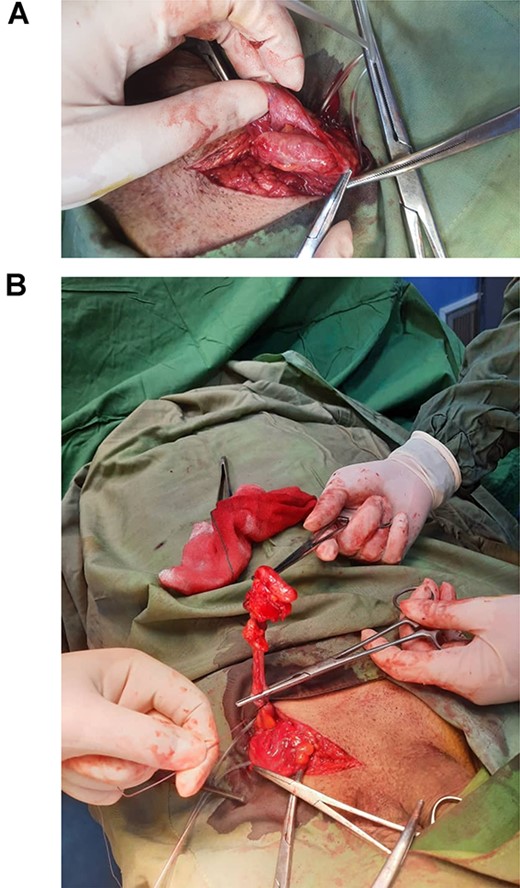
The patient went through spinal anesthesia under sterile conditions, the skin and subcutaneous in the right inguinal area were opened in the typical location. Afterward, the external fascia was opened and released, then the ilioinguinal and iliohypogastric nerves were explored and preserved, and the external ring was opened. The location of previous surgery was visible in the inguinal region. Chordae was released from the tuberculous pubic and cremasteric muscles were released by cautery and explored. Hernia sac was explored indirectly and released. Hernia sac was contained of inflamed appendix (Fig. 1A and B), then appendectomy was performed and the sample was sent for biopsy. The floor of inguinal canal was sutured by nylon yarn and repaired by Bassini method. His follow-up was done and he had no significant post-surgical complications.

(A) Inflamed appendix in inguinal sac (Amyand’s hernia). (B) Appendix extracted from inguinal hernia.
Pathological types of Amyand’s hernia and their management by Losanoff et al. [13]
| Type of hernia . | 1 . | 2 . | 3 . | 4 . |
|---|---|---|---|---|
| Salient features | Normal appendix | Acute appendicitis localized in the sac | Acute appendicitis, peritonitis | Acute appendicitis, other abdominal pathology |
| Surgical management | Reduction or appendectomy (depending on age), mesh hernioplasty | Appendectomy through hernia, endogenous repair | Appendectomy through laparotomy, endogenous repair | Appendectomy, diagnostic workup and other procedures as appropriate |
DISCUSSION
Inguinal hernia occurs mostly in two periods of time. One in childhood due to patent vaginalis process and again in elderly because of muscle weakness and dilation of inguinal canal. Inguinal hernia is one of the most common general surgeries worldwide [10]. Despite of high prevalence, Amyand’s hernia is a rare disease which cannot be diagnosed before the operation [11]. Inguinal hernia usually is simple to diagnosis and it makes pre-operative procedures such as ultrasonography (USG) and abdominal CT-scan unnecessary. Thus, specification of the organ involved in hernia sac pre-operatively is not routine and that is why Amyand’s hernia is an incidental finding during surgery [11, 12]. However, pre-operative USG was performed for this patient and it was considerable that intestinal loop stuck in hernia cavity was reported which during the operation showed no signs of intestinal loop in the hernia sac and also the patient’s history did not match the USG.
Amyand’s hernia is classified into four types. Type 1 is a normal, non-inflamed appendix in hernia sac, which is diagnosed only during operation incidentally, whereas type 2 is defined as inflamed appendix in hernia cavity. In this type, patient is ill and may have mild fever, abdominal tenderness, nausea or vomiting. These are the most common types of Amyand’s hernia (Table 1) [13]. It is interesting that in spite of long-term bulging and high pressure of appendix in hernia sac and obvious appendicitis on operation, our patient had no symptoms of fever, nausea, vomiting or abdominal tenderness and that may be due to neuropathy complication of patient’s diabetes or his mental illness [14, 15]. As the types progress, patient gets more complicated. In type 1 and 2 Amyand’s hernia, surgical approach is the same and contains of appendectomy along with herniorraphy. Due to abdominal wall infection it is not safe to use mesh in repair of contaminated abdominal wall, as in this operation we did not use mesh to repair abdominal wall after herniotomy [6, 13].
CONFLICT OF INTEREST STATEMENT
Authors disprove any financial support and had no conflict of interest to disclose.
References
- physical activity
- diabetes mellitus
- appendicitis
- physical examination
- ambulatory care facilities
- erythema
- hernias
- hernia, inguinal
- inguinal canal
- intestines
- pain
- abdomen
- diagnostic imaging
- incarcerated hernia
- simple excision of inguinal hernial sac
- incarcerated inguinal hernia
- causality
- abdominal fascia


