-
PDF
- Split View
-
Views
-
Cite
Cite
Mark Redden, Marjan Ghadiri, Acute appendicitis with associated trichobezoar of feline hair, Journal of Surgical Case Reports, Volume 2022, Issue 3, March 2022, rjac133, https://doi.org/10.1093/jscr/rjac133
Close - Share Icon Share
Abstract
A trichobezoar is an accumulation of ingested hair that forms a mass within the gastro-intestinal tract. Thrichobezoars usually consist of human hair and are known to cause obstruction and even perforation of gastrointestinal organs. There have been approximately seven reported cases of acute appendicitis with association trichobezoars found at the time of appendicectomy. We report a unique case of acute appendicitis with an associated trichobezoar of feline hair. A 15-year-old male presented with a 24-hour history of abdominal pain. A computed tomography scan demonstrated features of appendicitis with several hyperdensities within the base of the appendix. At the time of appendicectomy, the appendix was found to be perforated at the base. Faecoliths were identified containing numerous short, light-coloured hairs. Following the procedure, the family confirmed that they have a pet cat with short, light-coloured hair. The patient had an uneventful recovery.
INTRODUCTION
Acute appendicitis is a common cause of abdominal pain presenting to the emergency department. Appendicitis secondary to an appendiceal foreign body is a rare entity, which represents only 0.0005% of acute appendicitis cases [1]. Even rarer still is appendicitis due to a trichobezoar with only seven reported cases [1–7]. In these cases, the hair is usually human resulting from psychological conditions with trichotillomania (compulsive hair pulling) and trichophagia (compulsive hair swallowing) [2]. We present an unusual case of acute appendicitis secondary to a trichobezoar of ingested feline hair.
CASE
A 15-year-old male presented to the emergency department with a 24-hour history of abdominal pain and fevers. On examination, there was tenderness with guarding in the right lower quadrant of the abdomen. His vital signs were normal. The patient had no known medical conditions and had not had any previous surgery.
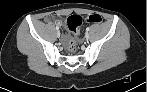
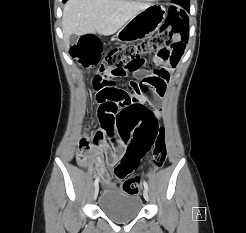
Investigations were arranged. His white cell count was 15.5 x 10⁹/L and C reactive protein was 117 mg/L. An ultrasound of the abdomen showed a small volume of free fluid in the right iliac fossa but did not identify the appendix. A computed tomography (CT) scan of the abdomen was performed, which showed that the appendix was dilated to 13 mm with associated peri-appendiceal fat stranding and several hyperdensities measuring up to 8 mm within the base of the appendix (Figs 1 and 2).
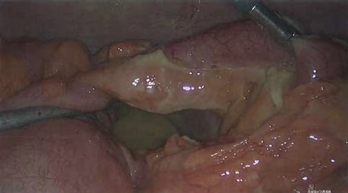
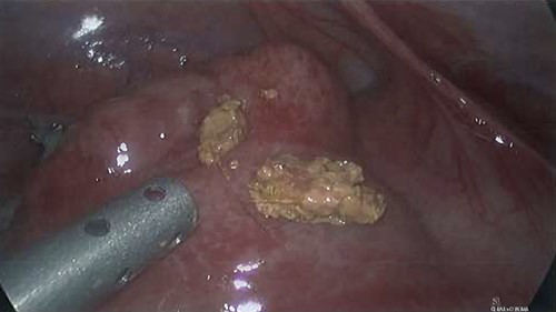
The patient proceeded to laparoscopic appendicectomy. At the outset of the procedure a cluster of short, firm, light coloured hairs were removed from the umbilicus. These hairs were different to the patient’s own and appeared like animal hair. Intra-operatively, the appendix was found to be inflamed with a perforated base (Fig. 3). Faecoliths, containing a large volume of hairs identical to those found in the umbilicus, had escaped the appendix through the perforation (Fig. 4). Pus was identified in all four quadrants of the abdomen. After dissection of the meso-appendix, a partial caecectomy was performed using a laparoscopic stapler. The hairs were carefully removed, and the abdomen was irrigated. A drain was placed in the pelvis.

Axial CT image demonstrating the inflamed appendix containing hyperdensities.

Coronal CT image demonstrating the inflamed appendix containing hyperdensities.


Following the procedure, the patient’s family confirmed that they have a pet cat with short light-coloured hair. The patient stayed in hospital for 3 days of intra-venous antibiotics. The drain was removed, and he was discharged with 5 days of oral antibiotics. He has since been reviewed in the outpatient clinic and has had an uneventful recovery.
DISCUSSION
Ingested foreign bodies are an accepted aetiology for acute appendicitis [8]. The first successful appendicectomy, which was performed in 1735, was for an ingested pin that had perforated the appendix. Common culprits throughout medical history have included metal pins and lead shot from game meat [8]. The work-up and management of these cases is usually identical to traditional appendicitis with the foreign body being an incidental finding on imaging, at the time of surgery or in the pathology laboratory.
A trichobezoar is an accumulation of ingested hair forming a mass within the gastro-intestinal tract, which can cause various issues. The most common complications of trichobezoars are perforation, intussusception and obstruction [7]. Rapunzel syndrome, which was first reported in 1968, is the presence of a gastric trichobezoar with a tail extending into the small bowel causing features of obstruction [9]. There is a presumed association with trichotillomania and trichophagia, however, this is frequently denied by the patients [9]. There has been approximately seven reported cases of trichobezoars being found at the time of appendicectomy for acute appendicitis [1–7]. It is suggested that the trichobezoars likely contributed to the development of appendicitis in these cases due to their location within the base of the appendixes.
This case is distinct from other cases of appendicitis with associated trichobezoars as the hair is likely of feline origin. There has only been one other reported case of animal hair causing pathology of the appendix [10]. Miller et al. reported a case of a 2-year-old boy who presented with signs and symptoms of appendicitis. At the time of appendicectomy, a canine hair was found to have perforated the tip of the appendix [10].
At this stage, defining the aetiology of a particular case of appendicitis has no bearing on the management of the individual patient. However, growing the shared knowledge of potential causes of appendicitis helps to provide a greater understanding of the disease process.
CONFLICT OF INTEREST STATEMENT
None declared.
FUNDING
None.


